{"title":"Placental Macrovascular Pattern from Pregnancies with Maternal Hypertensive and Fetal Growth Capacity Complications.","authors":"Kamilya Makhambetova, Yevgeniy Kamyshanskiy, Olga Ponamareva, Zhanna Amirbekova, Nazerke Oshakhtiyeva, Saule Kunanbaeva","doi":"10.3390/pathophysiology31040050","DOIUrl":null,"url":null,"abstract":"<p><p>Histomorphometric measurements of the wall thickness and internal diameter of the macrovessels of the chorionic villi of placentas from pregnancies complicated by preeclampsia or fetal growth restriction in comparison with normotensive pregnancy.</p><p><strong>Methods: </strong>The research included placentas from singleton pregnancies complicated by preeclampsia and/or fetal growth restriction, women delivered in medical institutions in Karaganda city (Kazakhstan). Placentas were divided into three groups: PE (<i>n</i> = 59), isolated FGR (<i>n</i> = 24), and PE with FGR (<i>n</i> = 41). The control group consisted of normotensive pregnancies, compared by gestation period. Placental examination and selection of placental tissue fragments were carried out in accordance with the consensus recommendations of the Amsterdam Placental Workshop Group. The sections were stained with hematoxylin and eosin and Masson trichrome. Morphometric measurements were performed using ImageJ software version 1.52p.</p><p><strong>Results: </strong>Our data showed that, in the PE group, there was a significant decrease in the wall thickness of the proximal and distal vessels with an increase in internal diameter compared with the control group (<i>p</i> < 0.01). In the PE + FGR group, there was a thickening of the wall of the proximal part of the vessels with a decrease in their lumen and a decrease in the wall thickness of the vessels with an increase in the lumen in the distal part compared with the control group (<i>p</i> < 0.01).</p><p><strong>Conclusions: </strong>Two histopatterns of placental macrovessels in preeclampsia were revealed: the histophenotype of diffuse (proximal and distal) ectatic macroangiopathy with a thin vascular wall with a decrease in the thickness of the muscle layer and the histophenotype of proximal fibromuscular sclerosis with vascular obliteration/spasm and distal ectatic macroangiopathy. We believe that significant structural differences in vascular remodeling may reflect the different temporal and spatial nature of the pathological factor. Future research is needed to investigate the associations between histopatterns of placental vascular remodeling in preeclampsia and long-term perinatal/maternal outcomes.</p>","PeriodicalId":19852,"journal":{"name":"Pathophysiology","volume":"31 4","pages":"699-708"},"PeriodicalIF":2.6000,"publicationDate":"2024-12-05","publicationTypes":"Journal Article","fieldsOfStudy":null,"isOpenAccess":false,"openAccessPdf":"https://www.ncbi.nlm.nih.gov/pmc/articles/PMC11679333/pdf/","citationCount":"0","resultStr":null,"platform":"Semanticscholar","paperid":null,"PeriodicalName":"Pathophysiology","FirstCategoryId":"1085","ListUrlMain":"https://doi.org/10.3390/pathophysiology31040050","RegionNum":0,"RegionCategory":null,"ArticlePicture":[],"TitleCN":null,"AbstractTextCN":null,"PMCID":null,"EPubDate":"","PubModel":"","JCR":"Q2","JCRName":"PATHOLOGY","Score":null,"Total":0}
引用次数: 0
Abstract
Histomorphometric measurements of the wall thickness and internal diameter of the macrovessels of the chorionic villi of placentas from pregnancies complicated by preeclampsia or fetal growth restriction in comparison with normotensive pregnancy.
Methods: The research included placentas from singleton pregnancies complicated by preeclampsia and/or fetal growth restriction, women delivered in medical institutions in Karaganda city (Kazakhstan). Placentas were divided into three groups: PE (n = 59), isolated FGR (n = 24), and PE with FGR (n = 41). The control group consisted of normotensive pregnancies, compared by gestation period. Placental examination and selection of placental tissue fragments were carried out in accordance with the consensus recommendations of the Amsterdam Placental Workshop Group. The sections were stained with hematoxylin and eosin and Masson trichrome. Morphometric measurements were performed using ImageJ software version 1.52p.
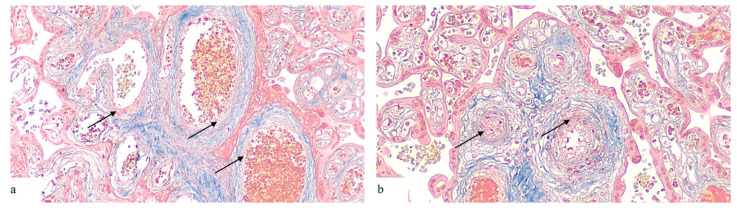
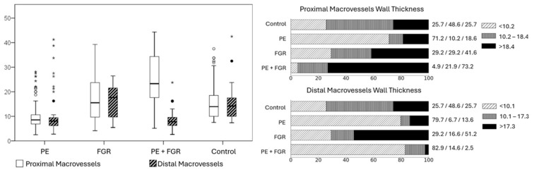
Results: Our data showed that, in the PE group, there was a significant decrease in the wall thickness of the proximal and distal vessels with an increase in internal diameter compared with the control group (p < 0.01). In the PE + FGR group, there was a thickening of the wall of the proximal part of the vessels with a decrease in their lumen and a decrease in the wall thickness of the vessels with an increase in the lumen in the distal part compared with the control group (p < 0.01).
Conclusions: Two histopatterns of placental macrovessels in preeclampsia were revealed: the histophenotype of diffuse (proximal and distal) ectatic macroangiopathy with a thin vascular wall with a decrease in the thickness of the muscle layer and the histophenotype of proximal fibromuscular sclerosis with vascular obliteration/spasm and distal ectatic macroangiopathy. We believe that significant structural differences in vascular remodeling may reflect the different temporal and spatial nature of the pathological factor. Future research is needed to investigate the associations between histopatterns of placental vascular remodeling in preeclampsia and long-term perinatal/maternal outcomes.
期刊介绍:
Pathophysiology is an international journal which publishes papers in English which address the etiology, development, and elimination of pathological processes. Contributions on the basic mechanisms underlying these processes, model systems and interdisciplinary approaches are strongly encouraged.


 分享
分享
 求助内容:
求助内容: 应助结果提醒方式:
 扫码关注我们
