Fully-automated segmentation of muscle and inter-/intra-muscular fat from magnetic resonance images of calves and thighs: an open-source workflow in Python.
Kenneth Tam, Si Wen Liu, Sarah Costa, Eva Szabo, Shannon Reitsma, Hana Gillick, Jonathan D Adachi, Andy Kin On Wong
{"title":"Fully-automated segmentation of muscle and inter-/intra-muscular fat from magnetic resonance images of calves and thighs: an open-source workflow in Python.","authors":"Kenneth Tam, Si Wen Liu, Sarah Costa, Eva Szabo, Shannon Reitsma, Hana Gillick, Jonathan D Adachi, Andy Kin On Wong","doi":"10.1186/s13395-024-00365-z","DOIUrl":null,"url":null,"abstract":"<p><strong>Background: </strong>INTER- and INTRAmuscular fat (IMF) is elevated in high metabolic states and can promote inflammation. While magnetic resonance imaging (MRI) excels in depicting IMF, the lack of reproducible tools prevents the ability to measure change and track intervention success.</p><p><strong>Methods: </strong>We detail an open-source fully-automated iterative threshold-seeking algorithm (ITSA) for segmenting IMF from T1-weighted MRI of the calf and thigh within three cohorts (CaMos Hamilton (N = 54), AMBERS (N = 280), OAI (N = 105)) selecting adults 45-85 years of age. Within the CaMos Hamilton cohort, same-day and 1-year repeated images (N = 38) were used to evaluate short- and long-term precision error with root mean square coefficients of variation; and to validate against semi-automated segmentation methods using linear regression. The effect of algorithmic improvements to fat ascertainment using 3D connectivity and partial volume correction rules on analytical precision was investigated. Robustness and versatility of the algorithm was demonstrated by application to different MR sequences/magnetic strength and to calf versus thigh scans.</p><p><strong>Results: </strong>Among 439 adults (319 female(89%), age: 71.6 ± 7.6 yrs, BMI: 28.06 ± 4.87 kg/m<sup>2</sup>, IMF%: 10.91 ± 4.57%), fully-automated ITSA performed well across MR sequences and anatomies from three cohorts. Applying both 3D connectivity and partial volume fat correction improved precision from 4.99% to 2.21% test-retest error. Validation against semi-automated methods showed R<sup>2</sup> from 0.92 to 0.98 with fully-automated ITSA routinely yielding more conservative computations of IMF volumes. Quality control shows 7% of cases requiring manual correction, primarily due to IMF merging with subcutaneous fat. A full workflow described methods to export tags for manual correction.</p><p><strong>Conclusions: </strong>The greatest challenge in segmenting IMF from MRI is in selecting a dynamic threshold that consistently performs across repeated imaging. Fully-automated ITSA achieved this, demonstrated low short- and long-term precision error, conducive of use within RCTs.</p>","PeriodicalId":21747,"journal":{"name":"Skeletal Muscle","volume":"14 1","pages":"37"},"PeriodicalIF":4.4000,"publicationDate":"2024-12-27","publicationTypes":"Journal Article","fieldsOfStudy":null,"isOpenAccess":false,"openAccessPdf":"https://www.ncbi.nlm.nih.gov/pmc/articles/PMC11674188/pdf/","citationCount":"0","resultStr":null,"platform":"Semanticscholar","paperid":null,"PeriodicalName":"Skeletal Muscle","FirstCategoryId":"3","ListUrlMain":"https://doi.org/10.1186/s13395-024-00365-z","RegionNum":2,"RegionCategory":"医学","ArticlePicture":[],"TitleCN":null,"AbstractTextCN":null,"PMCID":null,"EPubDate":"","PubModel":"","JCR":"Q2","JCRName":"CELL BIOLOGY","Score":null,"Total":0}
引用次数: 0
Abstract
Background: INTER- and INTRAmuscular fat (IMF) is elevated in high metabolic states and can promote inflammation. While magnetic resonance imaging (MRI) excels in depicting IMF, the lack of reproducible tools prevents the ability to measure change and track intervention success.
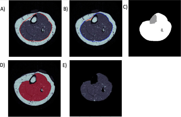
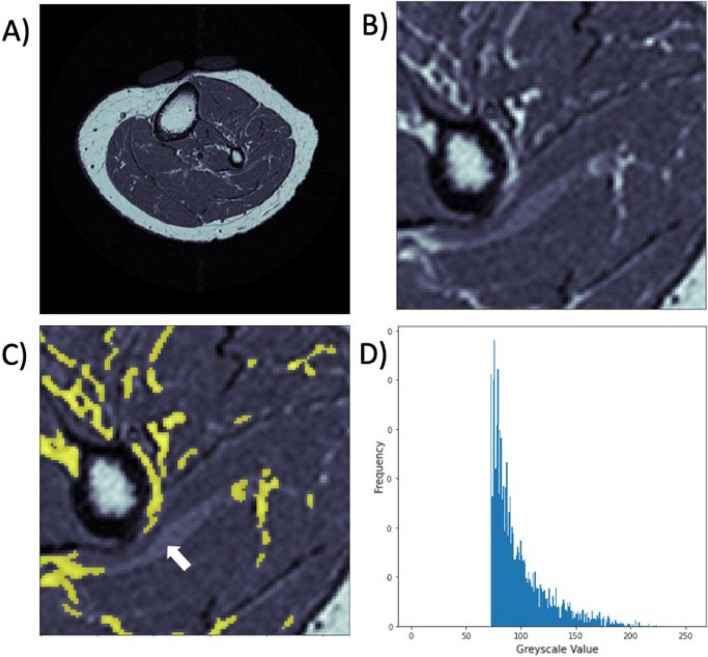
Methods: We detail an open-source fully-automated iterative threshold-seeking algorithm (ITSA) for segmenting IMF from T1-weighted MRI of the calf and thigh within three cohorts (CaMos Hamilton (N = 54), AMBERS (N = 280), OAI (N = 105)) selecting adults 45-85 years of age. Within the CaMos Hamilton cohort, same-day and 1-year repeated images (N = 38) were used to evaluate short- and long-term precision error with root mean square coefficients of variation; and to validate against semi-automated segmentation methods using linear regression. The effect of algorithmic improvements to fat ascertainment using 3D connectivity and partial volume correction rules on analytical precision was investigated. Robustness and versatility of the algorithm was demonstrated by application to different MR sequences/magnetic strength and to calf versus thigh scans.
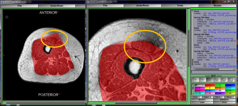
Results: Among 439 adults (319 female(89%), age: 71.6 ± 7.6 yrs, BMI: 28.06 ± 4.87 kg/m2, IMF%: 10.91 ± 4.57%), fully-automated ITSA performed well across MR sequences and anatomies from three cohorts. Applying both 3D connectivity and partial volume fat correction improved precision from 4.99% to 2.21% test-retest error. Validation against semi-automated methods showed R2 from 0.92 to 0.98 with fully-automated ITSA routinely yielding more conservative computations of IMF volumes. Quality control shows 7% of cases requiring manual correction, primarily due to IMF merging with subcutaneous fat. A full workflow described methods to export tags for manual correction.
Conclusions: The greatest challenge in segmenting IMF from MRI is in selecting a dynamic threshold that consistently performs across repeated imaging. Fully-automated ITSA achieved this, demonstrated low short- and long-term precision error, conducive of use within RCTs.
期刊介绍:
The only open access journal in its field, Skeletal Muscle publishes novel, cutting-edge research and technological advancements that investigate the molecular mechanisms underlying the biology of skeletal muscle. Reflecting the breadth of research in this area, the journal welcomes manuscripts about the development, metabolism, the regulation of mass and function, aging, degeneration, dystrophy and regeneration of skeletal muscle, with an emphasis on understanding adult skeletal muscle, its maintenance, and its interactions with non-muscle cell types and regulatory modulators.
Main areas of interest include:
-differentiation of skeletal muscle-
atrophy and hypertrophy of skeletal muscle-
aging of skeletal muscle-
regeneration and degeneration of skeletal muscle-
biology of satellite and satellite-like cells-
dystrophic degeneration of skeletal muscle-
energy and glucose homeostasis in skeletal muscle-
non-dystrophic genetic diseases of skeletal muscle, such as Spinal Muscular Atrophy and myopathies-
maintenance of neuromuscular junctions-
roles of ryanodine receptors and calcium signaling in skeletal muscle-
roles of nuclear receptors in skeletal muscle-
roles of GPCRs and GPCR signaling in skeletal muscle-
other relevant aspects of skeletal muscle biology.
In addition, articles on translational clinical studies that address molecular and cellular mechanisms of skeletal muscle will be published. Case reports are also encouraged for submission.
Skeletal Muscle reflects the breadth of research on skeletal muscle and bridges gaps between diverse areas of science for example cardiac cell biology and neurobiology, which share common features with respect to cell differentiation, excitatory membranes, cell-cell communication, and maintenance. Suitable articles are model and mechanism-driven, and apply statistical principles where appropriate; purely descriptive studies are of lesser interest.


 分享
分享
 求助内容:
求助内容: 应助结果提醒方式:
 扫码关注我们
