M-D Campoy, S Chiquillo-Enguix, V García-Sanz, J-C Pérez-Varela, S Camañes-Gonzalvo, V Paredes-Gallardo
{"title":"Is the mandibular buccal shelf anatomy related to craniofacial morphology? A cross-sectional CBCT study.","authors":"M-D Campoy, S Chiquillo-Enguix, V García-Sanz, J-C Pérez-Varela, S Camañes-Gonzalvo, V Paredes-Gallardo","doi":"10.4317/medoral.26897","DOIUrl":null,"url":null,"abstract":"<p><strong>Background: </strong>The placement of Temporary Anchorage Devices (TADs) in the mandibular buccal shelf area is a common option for distalizing the lower arch. Therefore, the study of bone thickness and depth in this area is mandatory before planning TAD insertion. The aim of this study was to quantify the width and depth of the mandibular buccal shelf structure and examine its associations with sex, age, skeletal class and vertical pattern.</p><p><strong>Material and methods: </strong>A cross-sectional study was carried out on cone beam computed tomographies obtained from 91 patients. The bone thickness was evaluated in the mandibular buccal shelf area 5 and 8 mm apical to the cement-enamel junction (CEJ), and the bone depth was measured 4 mm buccal to the CEJ at the level of the distal root of the mandibular first molar and the mesial root of the mandibular second molar using the InVivoDental 6.0 software.</p><p><strong>Results: </strong>The depth and thickness of the bone increased in distal areas, and the thickness was greater at 8 mm. No differences were found between sex or skeletal class. Bone thickness decreased with age, and it was significantly lower in hyperdivergent patients.</p><p><strong>Conclusions: </strong>The thickness of the bone was higher in distal and deeper areas, and the depth was greater in distal areas. The hyperdivergent facial pattern and age were negatively associated with bone thickness.</p>","PeriodicalId":49016,"journal":{"name":"Medicina Oral Patologia Oral Y Cirugia Bucal","volume":"30 1","pages":"e135-e140"},"PeriodicalIF":2.1000,"publicationDate":"2025-01-01","publicationTypes":"Journal Article","fieldsOfStudy":null,"isOpenAccess":false,"openAccessPdf":"https://www.ncbi.nlm.nih.gov/pmc/articles/PMC11801673/pdf/","citationCount":"0","resultStr":null,"platform":"Semanticscholar","paperid":null,"PeriodicalName":"Medicina Oral Patologia Oral Y Cirugia Bucal","FirstCategoryId":"3","ListUrlMain":"https://doi.org/10.4317/medoral.26897","RegionNum":3,"RegionCategory":"医学","ArticlePicture":[],"TitleCN":null,"AbstractTextCN":null,"PMCID":null,"EPubDate":"","PubModel":"","JCR":"Q2","JCRName":"DENTISTRY, ORAL SURGERY & MEDICINE","Score":null,"Total":0}
引用次数: 0
Abstract
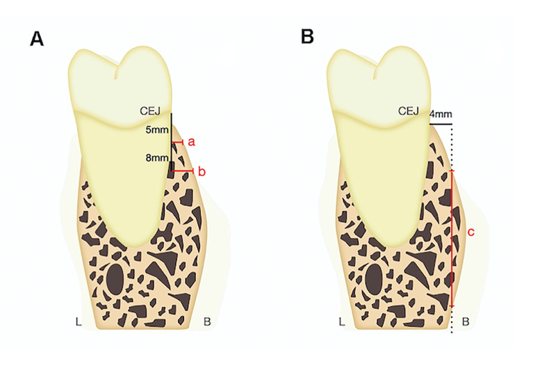
Background: The placement of Temporary Anchorage Devices (TADs) in the mandibular buccal shelf area is a common option for distalizing the lower arch. Therefore, the study of bone thickness and depth in this area is mandatory before planning TAD insertion. The aim of this study was to quantify the width and depth of the mandibular buccal shelf structure and examine its associations with sex, age, skeletal class and vertical pattern.
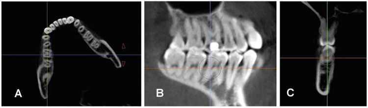
Material and methods: A cross-sectional study was carried out on cone beam computed tomographies obtained from 91 patients. The bone thickness was evaluated in the mandibular buccal shelf area 5 and 8 mm apical to the cement-enamel junction (CEJ), and the bone depth was measured 4 mm buccal to the CEJ at the level of the distal root of the mandibular first molar and the mesial root of the mandibular second molar using the InVivoDental 6.0 software.
Results: The depth and thickness of the bone increased in distal areas, and the thickness was greater at 8 mm. No differences were found between sex or skeletal class. Bone thickness decreased with age, and it was significantly lower in hyperdivergent patients.
Conclusions: The thickness of the bone was higher in distal and deeper areas, and the depth was greater in distal areas. The hyperdivergent facial pattern and age were negatively associated with bone thickness.
期刊介绍:
1. Oral Medicine and Pathology:
Clinicopathological as well as medical or surgical management aspects of
diseases affecting oral mucosa, salivary glands, maxillary bones, as well as
orofacial neurological disorders, and systemic conditions with an impact on
the oral cavity.
2. Oral Surgery:
Surgical management aspects of diseases affecting oral mucosa, salivary glands,
maxillary bones, teeth, implants, oral surgical procedures. Surgical management
of diseases affecting head and neck areas.
3. Medically compromised patients in Dentistry:
Articles discussing medical problems in Odontology will also be included, with
a special focus on the clinico-odontological management of medically compromised patients, and considerations regarding high-risk or disabled patients.
4. Implantology
5. Periodontology


 分享
分享
 求助内容:
求助内容: 应助结果提醒方式:
 扫码关注我们
