{"title":"Tuina alleviates the muscle atrophy induced by sciatic nerve injury in rats through regulation of PI3K/Akt signaling.","authors":"Yingqi Zhang, Hanyu Zhang, Jiayue Liu, Jiawei Sun, Yue Xu, Narentuya Shi, Hongzheng Zhang, Jiawang Yan, Jinping Chen, Hourong Wang, Tianyuan Yu","doi":"10.1186/s13018-024-05270-1","DOIUrl":null,"url":null,"abstract":"<p><strong>Background: </strong>Tuina is an effective treatment for the decrease of skeletal muscle atrophy after peripheral nerve injury. However, the underlying mechanism of action remains unclear. This study aimed to explore the underlying mechanisms of tuina in rats with sciatic nerve injury (SNI).</p><p><strong>Methods: </strong>We established an SNI rat model. After Tuina intervention, curative effects were evaluated by behavioral assessment, nerve function index, and muscle atrophy index (MAI). Pathological changes were observed by transmission electron microscopy and immunofluorescence. Insulin-like growth factor 1 (IGF-1), forkhead box O (FoxO) and p-FoxO levels were detected using enzyme-linked immunosorbent assay. Western blotting was performed to detect the expression of proteins involved in the PI3K/AKT signaling pathway.</p><p><strong>Result: </strong>Behavioral assessment, nerve function index, and MAI revealed that the tuina had significantly improved muscle atrophy after SNI compared with the SNI model group. Transmission electron microscopy showed that tuina improved muscle ultramicrostructure. CD31 immunofluorescence revealed that tuina improved microcirculation. Furthermore, we observed that tuina differentially regulated the levels of IGF-1, FoxO and p-FoxO, and the protein expression of p-Phosphoinositide 3-kinase (p-PI3K), p-AKT, and vascular endothelial growth factor in the anterior tibial muscle and soleus muscles.</p><p><strong>Conclusion: </strong>Tuina could effectively inhibit skeletal muscle atrophy via the microcirculation pathway in a rat model of SNI by regulating the expression of IGF-1 and FoxO. The underlying mechanism of action may involve the PI3K/Akt signaling pathway.</p>","PeriodicalId":16629,"journal":{"name":"Journal of Orthopaedic Surgery and Research","volume":"19 1","pages":"892"},"PeriodicalIF":2.8000,"publicationDate":"2024-12-31","publicationTypes":"Journal Article","fieldsOfStudy":null,"isOpenAccess":false,"openAccessPdf":"https://www.ncbi.nlm.nih.gov/pmc/articles/PMC11686904/pdf/","citationCount":"0","resultStr":null,"platform":"Semanticscholar","paperid":null,"PeriodicalName":"Journal of Orthopaedic Surgery and Research","FirstCategoryId":"3","ListUrlMain":"https://doi.org/10.1186/s13018-024-05270-1","RegionNum":3,"RegionCategory":"医学","ArticlePicture":[],"TitleCN":null,"AbstractTextCN":null,"PMCID":null,"EPubDate":"","PubModel":"","JCR":"Q1","JCRName":"ORTHOPEDICS","Score":null,"Total":0}
引用次数: 0
Abstract
Background: Tuina is an effective treatment for the decrease of skeletal muscle atrophy after peripheral nerve injury. However, the underlying mechanism of action remains unclear. This study aimed to explore the underlying mechanisms of tuina in rats with sciatic nerve injury (SNI).
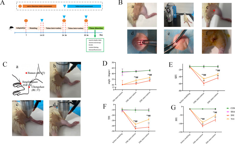
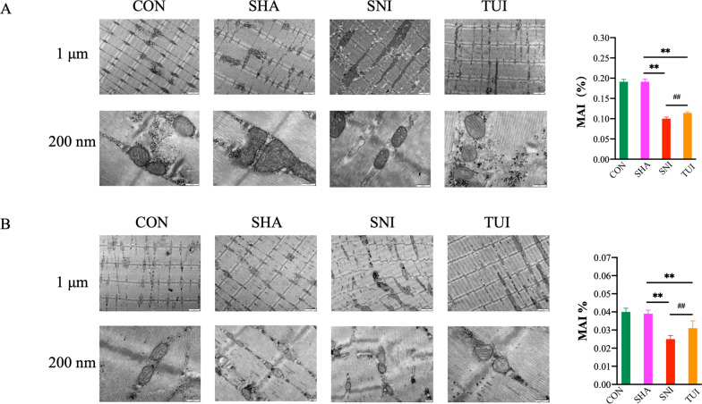
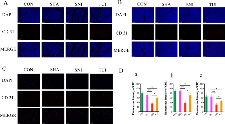
Methods: We established an SNI rat model. After Tuina intervention, curative effects were evaluated by behavioral assessment, nerve function index, and muscle atrophy index (MAI). Pathological changes were observed by transmission electron microscopy and immunofluorescence. Insulin-like growth factor 1 (IGF-1), forkhead box O (FoxO) and p-FoxO levels were detected using enzyme-linked immunosorbent assay. Western blotting was performed to detect the expression of proteins involved in the PI3K/AKT signaling pathway.
Result: Behavioral assessment, nerve function index, and MAI revealed that the tuina had significantly improved muscle atrophy after SNI compared with the SNI model group. Transmission electron microscopy showed that tuina improved muscle ultramicrostructure. CD31 immunofluorescence revealed that tuina improved microcirculation. Furthermore, we observed that tuina differentially regulated the levels of IGF-1, FoxO and p-FoxO, and the protein expression of p-Phosphoinositide 3-kinase (p-PI3K), p-AKT, and vascular endothelial growth factor in the anterior tibial muscle and soleus muscles.
Conclusion: Tuina could effectively inhibit skeletal muscle atrophy via the microcirculation pathway in a rat model of SNI by regulating the expression of IGF-1 and FoxO. The underlying mechanism of action may involve the PI3K/Akt signaling pathway.
期刊介绍:
Journal of Orthopaedic Surgery and Research is an open access journal that encompasses all aspects of clinical and basic research studies related to musculoskeletal issues.
Orthopaedic research is conducted at clinical and basic science levels. With the advancement of new technologies and the increasing expectation and demand from doctors and patients, we are witnessing an enormous growth in clinical orthopaedic research, particularly in the fields of traumatology, spinal surgery, joint replacement, sports medicine, musculoskeletal tumour management, hand microsurgery, foot and ankle surgery, paediatric orthopaedic, and orthopaedic rehabilitation. The involvement of basic science ranges from molecular, cellular, structural and functional perspectives to tissue engineering, gait analysis, automation and robotic surgery. Implant and biomaterial designs are new disciplines that complement clinical applications.
JOSR encourages the publication of multidisciplinary research with collaboration amongst clinicians and scientists from different disciplines, which will be the trend in the coming decades.


 分享
分享
 求助内容:
求助内容: 应助结果提醒方式:
 扫码关注我们
