Constanze Ramschütz, Nico Sollmann, Malek El Husseini, Karina Kupfer, Karolin J Paprottka, Maximilian T Löffler, Moritz R Hernandez Petzsche, Julian Schwarting, Jannis Bodden, Thomas Baum, Su Hwan Kim, Maria Wostrack, Claus Zimmer, Jan S Kirschke, Sebastian Rühling
{"title":"Cervicothoracic volumetric bone mineral density assessed by opportunistic QCT may be a reliable marker for osteoporosis in adults.","authors":"Constanze Ramschütz, Nico Sollmann, Malek El Husseini, Karina Kupfer, Karolin J Paprottka, Maximilian T Löffler, Moritz R Hernandez Petzsche, Julian Schwarting, Jannis Bodden, Thomas Baum, Su Hwan Kim, Maria Wostrack, Claus Zimmer, Jan S Kirschke, Sebastian Rühling","doi":"10.1007/s00198-024-07373-1","DOIUrl":null,"url":null,"abstract":"<p><p>This study aimed to validate the correlation between volumetric bone mineral density in the cervicothoracic and lumbar spine using measurements from opportunistic CT scans. The bone density assessment proved feasible, allowing us to propose optimal cut-off values for diagnosing osteoporosis and predicting vertebral fractures in the cervical and thoracic spine.</p><p><strong>Objectives: </strong>To investigate the performance of cervicothoracic volumetric bone mineral density (vBMD), obtained through opportunistic quantitative computed tomography (QCT), in discriminating patients with/without osteoporosis and with/without vertebral fractures (VFs), using lumbar vBMD as the reference.</p><p><strong>Methods: </strong>Three hundred twenty-five patients (65.3 ± 19.2 years, 140 women) with routine non-contrast or contrast-enhanced multi-detector CT (MDCT) scans were included. Trabecular vBMD was automatically extracted from each vertebra using a convolutional neural network (CNN)-based framework (SpineQ software v1.0) with asynchronous calibration and contrast phase correction. The correlations of vBMD between each vertebra spanning C2-T12 and the averaged lumbar spine (L1-L3, or L4 and L5) vBMD values were analyzed, considering fracture status and degeneration. Vertebra-specific linear regression equations were used to approximate lumbar vBMD at the cervicothoracic spine.</p><p><strong>Results: </strong>Cervicothoracic vBMD correlated well with lumbar vBMD (r = 0.79), with significant improvement after excluding degenerated vertebrae (p < 0.05; r = 0.89), except for C7-T3 and T9. Cervical (AUC = 0.94) and thoracic vBMD (AUC = 0.97) showed strong discriminatory ability for osteoporosis (vBMD < 80 mg/cm<sup>3</sup>). Excluding degenerated vertebrae at the cervical spine increased the AUC to 0.97. Cervical and thoracic vBMD (AUC = 0.74, AUC = 0.72) were comparable to lumbar vBMD (AUC = 0.72) in differentiating patients with and without prevalent VFs. Trabecular vBMD < 190 mg/cm<sup>3</sup> for the cervical spine and < 100 mg/cm<sup>3</sup> for the thoracic spine were potential indicators of osteoporosis, similar to < 80 mg/cm<sup>3</sup> at the lumbar spine.</p><p><strong>Conclusion: </strong>Cervicothoracic vBMD may allow for determination of osteoporosis and prediction of VFs.</p>","PeriodicalId":19638,"journal":{"name":"Osteoporosis International","volume":" ","pages":"423-433"},"PeriodicalIF":5.4000,"publicationDate":"2025-03-01","publicationTypes":"Journal Article","fieldsOfStudy":null,"isOpenAccess":false,"openAccessPdf":"https://www.ncbi.nlm.nih.gov/pmc/articles/PMC11882693/pdf/","citationCount":"0","resultStr":null,"platform":"Semanticscholar","paperid":null,"PeriodicalName":"Osteoporosis International","FirstCategoryId":"3","ListUrlMain":"https://doi.org/10.1007/s00198-024-07373-1","RegionNum":2,"RegionCategory":"医学","ArticlePicture":[],"TitleCN":null,"AbstractTextCN":null,"PMCID":null,"EPubDate":"2024/12/31 0:00:00","PubModel":"Epub","JCR":"Q1","JCRName":"ENDOCRINOLOGY & METABOLISM","Score":null,"Total":0}
引用次数: 0
Abstract
This study aimed to validate the correlation between volumetric bone mineral density in the cervicothoracic and lumbar spine using measurements from opportunistic CT scans. The bone density assessment proved feasible, allowing us to propose optimal cut-off values for diagnosing osteoporosis and predicting vertebral fractures in the cervical and thoracic spine.
Objectives: To investigate the performance of cervicothoracic volumetric bone mineral density (vBMD), obtained through opportunistic quantitative computed tomography (QCT), in discriminating patients with/without osteoporosis and with/without vertebral fractures (VFs), using lumbar vBMD as the reference.
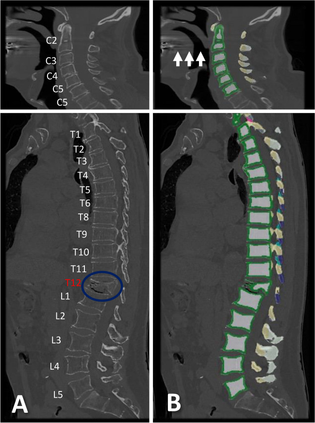
Methods: Three hundred twenty-five patients (65.3 ± 19.2 years, 140 women) with routine non-contrast or contrast-enhanced multi-detector CT (MDCT) scans were included. Trabecular vBMD was automatically extracted from each vertebra using a convolutional neural network (CNN)-based framework (SpineQ software v1.0) with asynchronous calibration and contrast phase correction. The correlations of vBMD between each vertebra spanning C2-T12 and the averaged lumbar spine (L1-L3, or L4 and L5) vBMD values were analyzed, considering fracture status and degeneration. Vertebra-specific linear regression equations were used to approximate lumbar vBMD at the cervicothoracic spine.
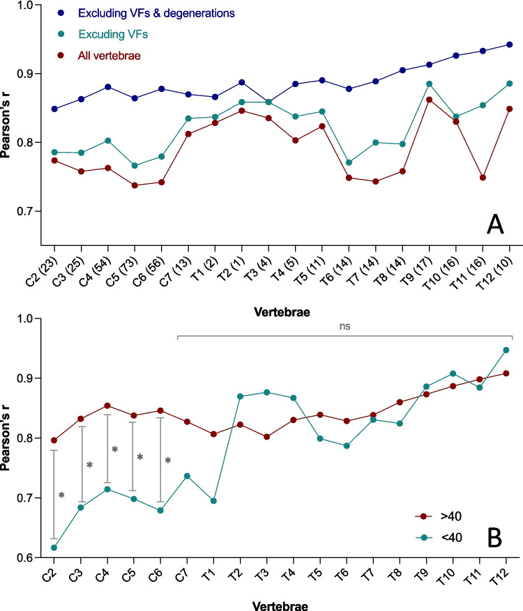
Results: Cervicothoracic vBMD correlated well with lumbar vBMD (r = 0.79), with significant improvement after excluding degenerated vertebrae (p < 0.05; r = 0.89), except for C7-T3 and T9. Cervical (AUC = 0.94) and thoracic vBMD (AUC = 0.97) showed strong discriminatory ability for osteoporosis (vBMD < 80 mg/cm3). Excluding degenerated vertebrae at the cervical spine increased the AUC to 0.97. Cervical and thoracic vBMD (AUC = 0.74, AUC = 0.72) were comparable to lumbar vBMD (AUC = 0.72) in differentiating patients with and without prevalent VFs. Trabecular vBMD < 190 mg/cm3 for the cervical spine and < 100 mg/cm3 for the thoracic spine were potential indicators of osteoporosis, similar to < 80 mg/cm3 at the lumbar spine.
Conclusion: Cervicothoracic vBMD may allow for determination of osteoporosis and prediction of VFs.
期刊介绍:
An international multi-disciplinary journal which is a joint initiative between the International Osteoporosis Foundation and the National Osteoporosis Foundation of the USA, Osteoporosis International provides a forum for the communication and exchange of current ideas concerning the diagnosis, prevention, treatment and management of osteoporosis and other metabolic bone diseases.
It publishes: original papers - reporting progress and results in all areas of osteoporosis and its related fields; review articles - reflecting the present state of knowledge in special areas of summarizing limited themes in which discussion has led to clearly defined conclusions; educational articles - giving information on the progress of a topic of particular interest; case reports - of uncommon or interesting presentations of the condition.
While focusing on clinical research, the Journal will also accept submissions on more basic aspects of research, where they are considered by the editors to be relevant to the human disease spectrum.


 分享
分享
 求助内容:
求助内容: 应助结果提醒方式:
 扫码关注我们
