Short-Term Hormonal Changes Following Microdissection Testicular Sperm Extraction among Men with Non-Obstructive Azoospermia: Findings from a Large Longitudinal Prospective Multicentric Study.
Fausto Negri, Edoardo Pozzi, Christian Corsini, Massimiliano Raffo, Federico Belladelli, Alessandro Bertini, Francesco Cattafi, Eugenio Ventimiglia, Rayan Matloob, Antonino Saccà, Luca Boeri, Alessia d'Arma, Francesco Montorsi, Andrea Salonia
{"title":"Short-Term Hormonal Changes Following Microdissection Testicular Sperm Extraction among Men with Non-Obstructive Azoospermia: Findings from a Large Longitudinal Prospective Multicentric Study.","authors":"Fausto Negri, Edoardo Pozzi, Christian Corsini, Massimiliano Raffo, Federico Belladelli, Alessandro Bertini, Francesco Cattafi, Eugenio Ventimiglia, Rayan Matloob, Antonino Saccà, Luca Boeri, Alessia d'Arma, Francesco Montorsi, Andrea Salonia","doi":"10.5534/wjmh.240184","DOIUrl":null,"url":null,"abstract":"<p><strong>Purpose: </strong>We aimed to investigate possible hormonal changes following microdissection testicular sperm extraction (mTESE) in men with non-obstructive azoospermia (NOA) across three referral centers.</p><p><strong>Materials and methods: </strong>We prospectively analyzed data from 102 consecutive NOA men. Patients with prior hormonal therapies were excluded. Preoperative serum hormone levels (total testosterone [tT], luteinizing hormone [LH], follicle-stimulating hormone [FSH], and 17β-estradiol) were collected, with repeat measurements at 3-month post-surgery. We divided the cohort into two groups using a tT cut-off value of 3 ng/mL: 1) men who kept eugonadal status; and, 2) men who were initially eugonadal but became testosterone deficient (TD) after surgery.</p><p><strong>Results: </strong>Overall, median (interquartile range [IQR]) age was 37 years (32-40 years). Positive sperm retrieval during mTESE was observed in 34 (33.3%) patients, and 48 (47.1%) underwent bilateral mTESE. Compared to baseline, 3-month postoperative median (IQR) hormonal levels were as follows: tT: 3.71 ng/mL (2.76-5.24 ng/mL) <i>vs.</i> 4.27 ng/mL (3.25-6.07 ng/mL), p=0.32; FSH: 22.0 mIU/mL (12.65-31.47 mIU/mL) <i>vs.</i> 19.5 mIU/mL (11.63-25.8 mIU/mL), p=0.25; LH: 9.0 mIU/mL (5.11-12.4 mIU/mL) <i>vs.</i> 7.6 mIU/mL (5.04-13.4 mIU/mL), p=0.73, respectively. Twelve (13.5%) eugonadal men at baseline showed TD after mTESE. Median (IQR) tT values at baseline and 3-month follow-up were compared between those who preserved eugonadal status after surgery and those who became TD after surgery: baseline levels were 4.46 ng/mL (4.1-6.27 ng/mL) <i>vs.</i> 4.14 ng/mL (3.24-4.98 ng/mL), p=0.09; and, 3-month follow-up levels were 4.58 ng/mL (3.58-5.56 ng/mL) <i>vs.</i> 2.51 ng/mL (2.31-2.76 ng/mL), p<0.001, respectively. Men who developed TD had lower testicular volume (TV) (6 [4-10] vs. 10 [8-12.25] Prader, p=0.001) and karyotype abnormalities (4 [33.3] <i>vs.</i> 1 [1.3], p=0.006).</p><p><strong>Conclusions: </strong>This multicentric study shows that mTESE in men with NOA does not significantly impact short-time postoperative follow-up tT, LH, and FSH levels. A substantial proportion of men who were initially eugonadal demonstrated tT suggestive for TD at 3-month follow-up. These men had lower TV at baseline and abnormal karyotype.</p>","PeriodicalId":54261,"journal":{"name":"World Journal of Mens Health","volume":" ","pages":"702-709"},"PeriodicalIF":4.1000,"publicationDate":"2025-07-01","publicationTypes":"Journal Article","fieldsOfStudy":null,"isOpenAccess":false,"openAccessPdf":"https://www.ncbi.nlm.nih.gov/pmc/articles/PMC12257323/pdf/","citationCount":"0","resultStr":null,"platform":"Semanticscholar","paperid":null,"PeriodicalName":"World Journal of Mens Health","FirstCategoryId":"3","ListUrlMain":"https://doi.org/10.5534/wjmh.240184","RegionNum":3,"RegionCategory":"医学","ArticlePicture":[],"TitleCN":null,"AbstractTextCN":null,"PMCID":null,"EPubDate":"2024/11/27 0:00:00","PubModel":"Epub","JCR":"Q1","JCRName":"ANDROLOGY","Score":null,"Total":0}
引用次数: 0
Abstract
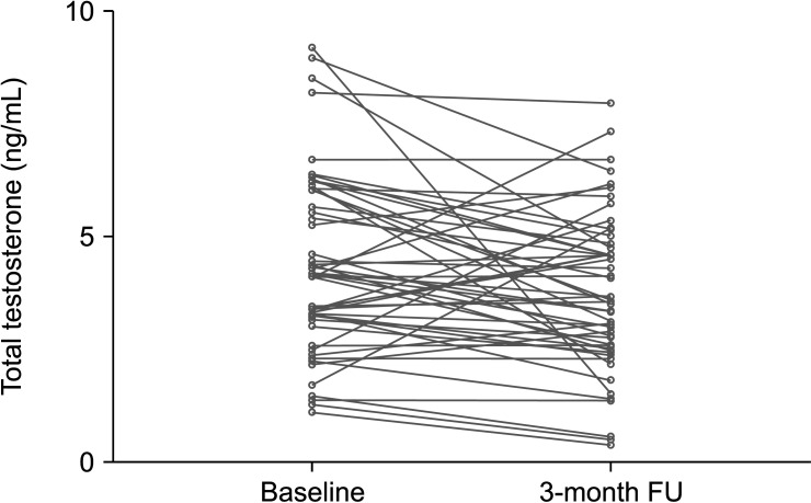
Purpose: We aimed to investigate possible hormonal changes following microdissection testicular sperm extraction (mTESE) in men with non-obstructive azoospermia (NOA) across three referral centers.
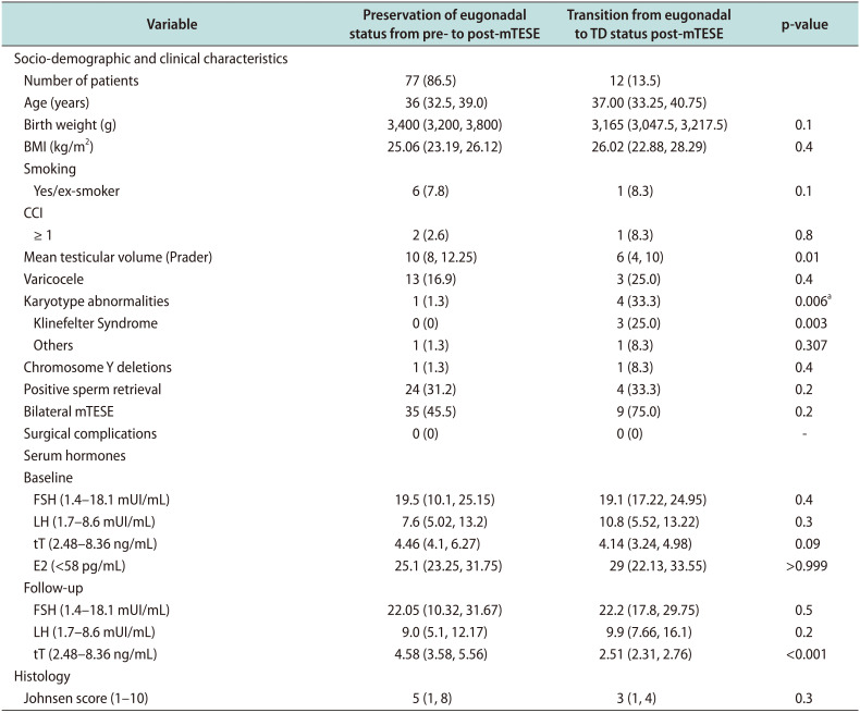
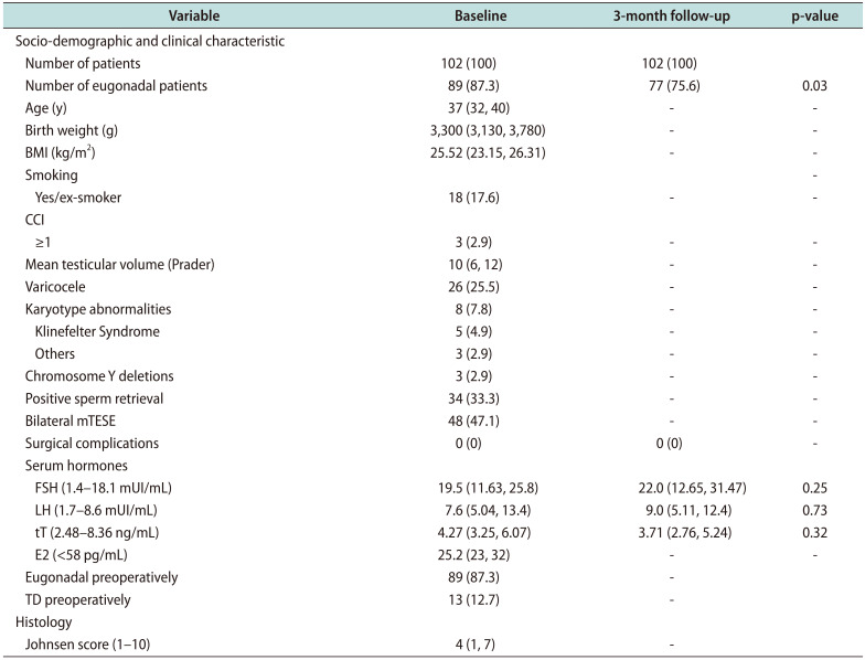
Materials and methods: We prospectively analyzed data from 102 consecutive NOA men. Patients with prior hormonal therapies were excluded. Preoperative serum hormone levels (total testosterone [tT], luteinizing hormone [LH], follicle-stimulating hormone [FSH], and 17β-estradiol) were collected, with repeat measurements at 3-month post-surgery. We divided the cohort into two groups using a tT cut-off value of 3 ng/mL: 1) men who kept eugonadal status; and, 2) men who were initially eugonadal but became testosterone deficient (TD) after surgery.
Results: Overall, median (interquartile range [IQR]) age was 37 years (32-40 years). Positive sperm retrieval during mTESE was observed in 34 (33.3%) patients, and 48 (47.1%) underwent bilateral mTESE. Compared to baseline, 3-month postoperative median (IQR) hormonal levels were as follows: tT: 3.71 ng/mL (2.76-5.24 ng/mL) vs. 4.27 ng/mL (3.25-6.07 ng/mL), p=0.32; FSH: 22.0 mIU/mL (12.65-31.47 mIU/mL) vs. 19.5 mIU/mL (11.63-25.8 mIU/mL), p=0.25; LH: 9.0 mIU/mL (5.11-12.4 mIU/mL) vs. 7.6 mIU/mL (5.04-13.4 mIU/mL), p=0.73, respectively. Twelve (13.5%) eugonadal men at baseline showed TD after mTESE. Median (IQR) tT values at baseline and 3-month follow-up were compared between those who preserved eugonadal status after surgery and those who became TD after surgery: baseline levels were 4.46 ng/mL (4.1-6.27 ng/mL) vs. 4.14 ng/mL (3.24-4.98 ng/mL), p=0.09; and, 3-month follow-up levels were 4.58 ng/mL (3.58-5.56 ng/mL) vs. 2.51 ng/mL (2.31-2.76 ng/mL), p<0.001, respectively. Men who developed TD had lower testicular volume (TV) (6 [4-10] vs. 10 [8-12.25] Prader, p=0.001) and karyotype abnormalities (4 [33.3] vs. 1 [1.3], p=0.006).
Conclusions: This multicentric study shows that mTESE in men with NOA does not significantly impact short-time postoperative follow-up tT, LH, and FSH levels. A substantial proportion of men who were initially eugonadal demonstrated tT suggestive for TD at 3-month follow-up. These men had lower TV at baseline and abnormal karyotype.


 分享
分享
 求助内容:
求助内容: 应助结果提醒方式:
 扫码关注我们
