Rosai-Dorfman Disease Presenting With FDG-Avid Breast Masses and Axillary Lymph Nodes on PET-CT in a Patient With Recent Diagnosis of Endometrial Carcinoma: A Diagnostic Dilemma.
Jennifer Kudja-Rennick, Pai Raghav, Cedric Pluguez-Turull, Katherine Drews-Elger, Cristina Hoyos
{"title":"Rosai-Dorfman Disease Presenting With FDG-Avid Breast Masses and Axillary Lymph Nodes on PET-CT in a Patient With Recent Diagnosis of Endometrial Carcinoma: A Diagnostic Dilemma.","authors":"Jennifer Kudja-Rennick, Pai Raghav, Cedric Pluguez-Turull, Katherine Drews-Elger, Cristina Hoyos","doi":"10.4274/ejbh.galenos.2024.2024-9-2","DOIUrl":null,"url":null,"abstract":"<p><p>Rosai-Dorfman disease (RDD) is a self-limited, idiopathic, non-neoplastic disorder characterized by the proliferation of phagocytic histiocytes, which can mimic malignant lymphoproliferative disease. Cases of RDD most commonly present as bilateral painless cervical lymphadenopathy, with lesser involvement of the axilla, inguinal, and mediastinal lymph nodes. We present the case of a 62-year-old woman with a history of endometrial serous carcinoma who underwent evaluation at a dedicated breast imaging department after positron emission tomography/computed tomography (PET/CT) revealed breast masses and axillary nodes with increased uptake of fluorodeoxyglucose (FDG). Upon clinical examination, she had bilateral palpable lumps in both breasts and axillae. Subsequent dedicated breast imaging with bilateral diagnostic mammography with tomosynthesis and bilateral complete breast ultrasound were suspicious for malignancy detecting bilateral breast masses and axillary lymphadenopathy corresponding to the FDG-avid findings on PET/CT. Ultrasound-guided core needle biopsies, however, revealed a diagnosis of RDD. This case highlights the unique characteristics of RDD with an atypical clinical presentation suspicious for breast cancer both clinically and radiologically.</p>","PeriodicalId":93996,"journal":{"name":"European journal of breast health","volume":"21 1","pages":"74-79"},"PeriodicalIF":1.7000,"publicationDate":"2025-01-01","publicationTypes":"Journal Article","fieldsOfStudy":null,"isOpenAccess":false,"openAccessPdf":"https://www.ncbi.nlm.nih.gov/pmc/articles/PMC11706118/pdf/","citationCount":"0","resultStr":null,"platform":"Semanticscholar","paperid":null,"PeriodicalName":"European journal of breast health","FirstCategoryId":"1085","ListUrlMain":"https://doi.org/10.4274/ejbh.galenos.2024.2024-9-2","RegionNum":0,"RegionCategory":null,"ArticlePicture":[],"TitleCN":null,"AbstractTextCN":null,"PMCID":null,"EPubDate":"","PubModel":"","JCR":"Q4","JCRName":"ONCOLOGY","Score":null,"Total":0}
引用次数: 0
Abstract
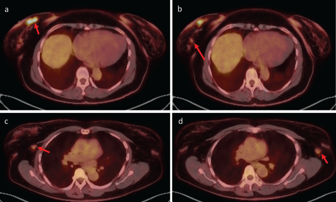
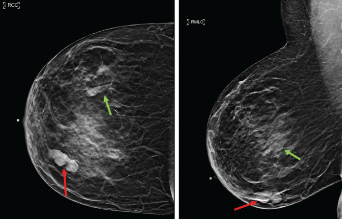
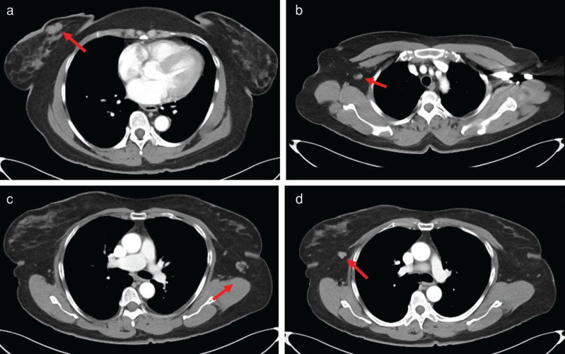
Rosai-Dorfman disease (RDD) is a self-limited, idiopathic, non-neoplastic disorder characterized by the proliferation of phagocytic histiocytes, which can mimic malignant lymphoproliferative disease. Cases of RDD most commonly present as bilateral painless cervical lymphadenopathy, with lesser involvement of the axilla, inguinal, and mediastinal lymph nodes. We present the case of a 62-year-old woman with a history of endometrial serous carcinoma who underwent evaluation at a dedicated breast imaging department after positron emission tomography/computed tomography (PET/CT) revealed breast masses and axillary nodes with increased uptake of fluorodeoxyglucose (FDG). Upon clinical examination, she had bilateral palpable lumps in both breasts and axillae. Subsequent dedicated breast imaging with bilateral diagnostic mammography with tomosynthesis and bilateral complete breast ultrasound were suspicious for malignancy detecting bilateral breast masses and axillary lymphadenopathy corresponding to the FDG-avid findings on PET/CT. Ultrasound-guided core needle biopsies, however, revealed a diagnosis of RDD. This case highlights the unique characteristics of RDD with an atypical clinical presentation suspicious for breast cancer both clinically and radiologically.


 分享
分享

 求助内容:
求助内容: 应助结果提醒方式:
 扫码关注我们
