Omid Moghaddas, Ehsan Seyedjafari, Donya Sadat Mahoutchi
{"title":"Biological behavior of mesenchymal stem cells on two types of commercial dermal scaffolds: An in vitro study.","authors":"Omid Moghaddas, Ehsan Seyedjafari, Donya Sadat Mahoutchi","doi":"10.34172/japid.2024.015","DOIUrl":null,"url":null,"abstract":"<p><strong>Background: </strong>Acellular dermal matrix (ADM) has been introduced as an alternative to autogenous grafts. This study assessed the biological behavior of mesenchymal stem cells (MSCs) on two types of commercial ADM scaffolds.</p><p><strong>Methods: </strong>The present in vitro study investigated the behavior of MSCs cultured on scaffold type I CenoDerm® (Tissue Regeneration Corporation) and type II Acellular Dermis (Iranian Tissue Product Co.) as the test groups and an empty well plate as the control group (n=78). Cell attachment was assessed after 12 hours of incubation using 6,4-diamidino-2-phenylindole (DAPI) staining and methyl thiazole tetrazolium (MTT) assay. Cell proliferation was assessed using the MTT assay at 24- and 84-hour and 7-day intervals. Cell morphology was also assessed under a scanning electron microscope (SEM) at 24 hours. MTT assay and DAPI staining were repeated for five samples in all the three groups. Mann-Whitney, ANOVA, and post hoc Tukey tests were used for statistical analysis.</p><p><strong>Results: </strong>The DAPI staining and MTT assay showed similar results concerning cell attachment between all the groups at 12 hours (<i>P</i>=0.4). At 24 hours, cell proliferation was significantly higher in scaffold groups (<i>P</i><0.001). At seven days, the lowest cell proliferation was noted in the scaffold II group, with a significant difference between the groups (<i>P</i>=0.01). At 24 hours, cell expansion was greater in the control group, followed by the scaffold I group.</p><p><strong>Conclusion: </strong>Both scaffolds were similar in MSC attachment, but scaffold I appeared superior to scaffold II in terms of MSC proliferation and morphology in vitro.</p>","PeriodicalId":73584,"journal":{"name":"Journal of advanced periodontology & implant dentistry","volume":"16 2","pages":"133-138"},"PeriodicalIF":0.0000,"publicationDate":"2024-08-11","publicationTypes":"Journal Article","fieldsOfStudy":null,"isOpenAccess":false,"openAccessPdf":"https://www.ncbi.nlm.nih.gov/pmc/articles/PMC11699267/pdf/","citationCount":"0","resultStr":null,"platform":"Semanticscholar","paperid":null,"PeriodicalName":"Journal of advanced periodontology & implant dentistry","FirstCategoryId":"1085","ListUrlMain":"https://doi.org/10.34172/japid.2024.015","RegionNum":0,"RegionCategory":null,"ArticlePicture":[],"TitleCN":null,"AbstractTextCN":null,"PMCID":null,"EPubDate":"2024/1/1 0:00:00","PubModel":"eCollection","JCR":"","JCRName":"","Score":null,"Total":0}
引用次数: 0
Abstract
Background: Acellular dermal matrix (ADM) has been introduced as an alternative to autogenous grafts. This study assessed the biological behavior of mesenchymal stem cells (MSCs) on two types of commercial ADM scaffolds.
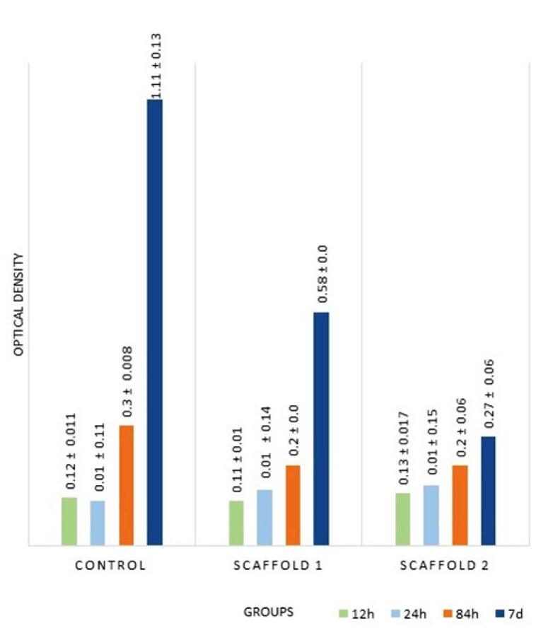
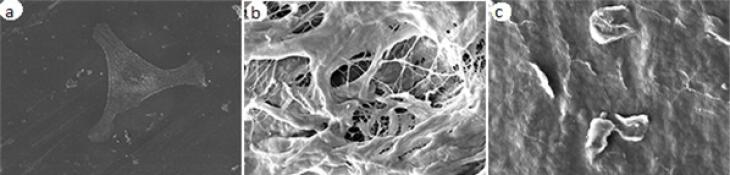
Methods: The present in vitro study investigated the behavior of MSCs cultured on scaffold type I CenoDerm® (Tissue Regeneration Corporation) and type II Acellular Dermis (Iranian Tissue Product Co.) as the test groups and an empty well plate as the control group (n=78). Cell attachment was assessed after 12 hours of incubation using 6,4-diamidino-2-phenylindole (DAPI) staining and methyl thiazole tetrazolium (MTT) assay. Cell proliferation was assessed using the MTT assay at 24- and 84-hour and 7-day intervals. Cell morphology was also assessed under a scanning electron microscope (SEM) at 24 hours. MTT assay and DAPI staining were repeated for five samples in all the three groups. Mann-Whitney, ANOVA, and post hoc Tukey tests were used for statistical analysis.
Results: The DAPI staining and MTT assay showed similar results concerning cell attachment between all the groups at 12 hours (P=0.4). At 24 hours, cell proliferation was significantly higher in scaffold groups (P<0.001). At seven days, the lowest cell proliferation was noted in the scaffold II group, with a significant difference between the groups (P=0.01). At 24 hours, cell expansion was greater in the control group, followed by the scaffold I group.
Conclusion: Both scaffolds were similar in MSC attachment, but scaffold I appeared superior to scaffold II in terms of MSC proliferation and morphology in vitro.


 分享
分享

 求助内容:
求助内容: 应助结果提醒方式:
 扫码关注我们
