{"title":"A Rare Case of Adrenal Gland Metastasis from Parotid Adenocarcinoma: Unveiling the Potential Augmented Utility of FAPI PET/CT.","authors":"Akram Al-Ibraheem, Marwah Abdulrahman, Medyan Alrousan, Mohamad Haidar","doi":"10.4103/ijnm.ijnm_13_24","DOIUrl":null,"url":null,"abstract":"<p><p>Conventional imaging techniques, while essential, occasionally fall short in identifying elusive metastatic lesions, leading to delayed diagnoses and compromised patient outcomes. Gallium-68 fibroblast activating protein inhibitor (<sup>68</sup>Ga-FAPI) positron emission tomography/computed tomography (PET/CT), leveraging the distinct affinity of fibroblast activation protein for cancer-associated fibroblasts, emerges as a promising solution to bridge this diagnostic gap. Parotid gland adenocarcinoma is a relatively rare malignancy with metastasis typically occurring in regional lymph nodes and distant sites such as the lungs and bones. However, there have been limited reported cases of rare metastatic sites such as the adrenal gland. This exceptional case report details the clinical presentation, diagnostic workup, and management steps of a rare case of a 47-year-old female patient diagnosed with parotid gland adenocarcinoma with confusing metastasis to the ipsilateral adrenal gland which was confirmed later with a follow-up <sup>68</sup>Ga-FAPI PET/CT scan. We aim to highlight FAPI unique ability to illuminate metastatic foci in challenging anatomical locations.</p>","PeriodicalId":45830,"journal":{"name":"Indian Journal of Nuclear Medicine","volume":"39 4","pages":"309-312"},"PeriodicalIF":0.5000,"publicationDate":"2024-07-01","publicationTypes":"Journal Article","fieldsOfStudy":null,"isOpenAccess":false,"openAccessPdf":"https://www.ncbi.nlm.nih.gov/pmc/articles/PMC11708800/pdf/","citationCount":"0","resultStr":null,"platform":"Semanticscholar","paperid":null,"PeriodicalName":"Indian Journal of Nuclear Medicine","FirstCategoryId":"1085","ListUrlMain":"https://doi.org/10.4103/ijnm.ijnm_13_24","RegionNum":0,"RegionCategory":null,"ArticlePicture":[],"TitleCN":null,"AbstractTextCN":null,"PMCID":null,"EPubDate":"2024/11/18 0:00:00","PubModel":"Epub","JCR":"Q4","JCRName":"RADIOLOGY, NUCLEAR MEDICINE & MEDICAL IMAGING","Score":null,"Total":0}
引用次数: 0
Abstract
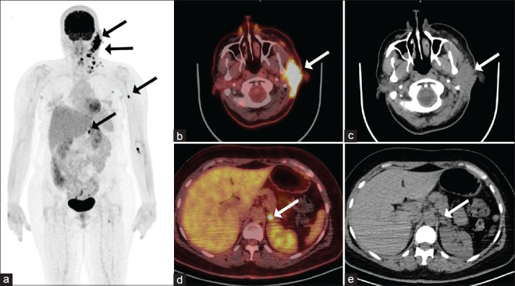
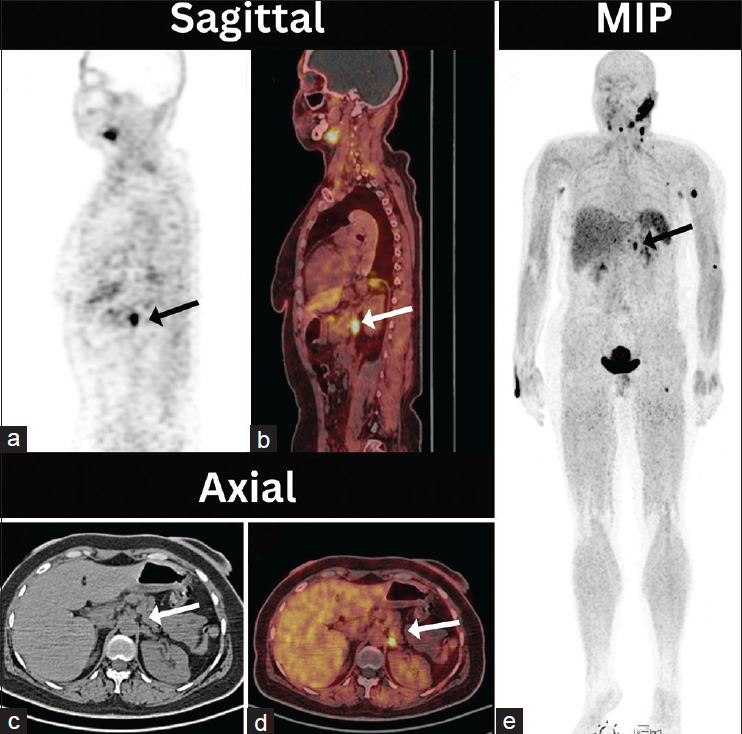
Conventional imaging techniques, while essential, occasionally fall short in identifying elusive metastatic lesions, leading to delayed diagnoses and compromised patient outcomes. Gallium-68 fibroblast activating protein inhibitor (68Ga-FAPI) positron emission tomography/computed tomography (PET/CT), leveraging the distinct affinity of fibroblast activation protein for cancer-associated fibroblasts, emerges as a promising solution to bridge this diagnostic gap. Parotid gland adenocarcinoma is a relatively rare malignancy with metastasis typically occurring in regional lymph nodes and distant sites such as the lungs and bones. However, there have been limited reported cases of rare metastatic sites such as the adrenal gland. This exceptional case report details the clinical presentation, diagnostic workup, and management steps of a rare case of a 47-year-old female patient diagnosed with parotid gland adenocarcinoma with confusing metastasis to the ipsilateral adrenal gland which was confirmed later with a follow-up 68Ga-FAPI PET/CT scan. We aim to highlight FAPI unique ability to illuminate metastatic foci in challenging anatomical locations.


 分享
分享
 求助内容:
求助内容: 应助结果提醒方式:
 扫码关注我们
