Nao Miyamae, Yuko Imakata, Mao Kunimitsu, Makoto Oe
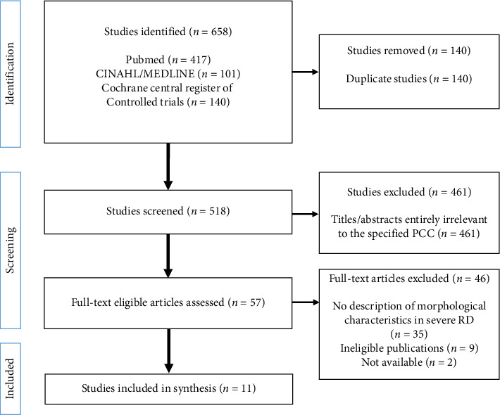
{"title":"Development and Healing Process of Severe Radiodermatitis in Patients With Head and Neck Cancer Undergoing Radiotherapy: A Scoping Review.","authors":"Nao Miyamae, Yuko Imakata, Mao Kunimitsu, Makoto Oe","doi":"10.1155/nrp/1940552","DOIUrl":null,"url":null,"abstract":"<p><p><b>Aims:</b> To summarize the morphological characteristics and development and healing processes of severe radiodermatitis for examining the factors contributing to the development of severe radiodermatitis in patients with head and neck cancer. <b>Methods:</b> This scoping review was conducted in accordance with PRISMA extension for Scoping Reviews. Data were extracted from selected references describing detailed conditions of severe radiodermatitis in patients with head and neck cancer. The data were organized separately for radiotherapy, chemoradiotherapy, and bioradiotherapy. <b>Data Sources:</b> Medline, PubMed, CINAHL, and Cochrane Central Register of Controlled Trials databases were used to search for papers from 2000 to December 2023. <b>Results:</b> 11 out of 658 references met the criteria for this review. The morphological characteristics of severe radiodermatitis were categorized by symptoms, site, and shape, and a condition in which moist desquamations and associated crusts spreading to the anterior and lateral neck areas were extracted. In bioradiotherapy, the process of keratinocyte degeneration and formation of blisters under the epidermis leading to moist desquamations was extracted. In chemoradiotherapy, the process of epithelization was extracted 1 week following the occurrence of moist desquamations. <b>Conclusions:</b> Moist desquamations are more likely to occur in severe radiodermatitis in patients with head and neck cancer. Since they can fuse and spread, preventative measures to mitigate spreading are important. However, there is insufficient information to examine the causes of widespread moist desquamations. For preventing moist desquamations and establishing care methods to heal moist desquamations, it may be necessary to identify the symptoms, site, and shape, including the color tone and depth, and healing process during their occurrence.</p>","PeriodicalId":46917,"journal":{"name":"Nursing Research and Practice","volume":"2024 ","pages":"1940552"},"PeriodicalIF":2.3000,"publicationDate":"2024-12-31","publicationTypes":"Journal Article","fieldsOfStudy":null,"isOpenAccess":false,"openAccessPdf":"https://www.ncbi.nlm.nih.gov/pmc/articles/PMC11707061/pdf/","citationCount":"0","resultStr":null,"platform":"Semanticscholar","paperid":null,"PeriodicalName":"Nursing Research and Practice","FirstCategoryId":"1085","ListUrlMain":"https://doi.org/10.1155/nrp/1940552","RegionNum":0,"RegionCategory":null,"ArticlePicture":[],"TitleCN":null,"AbstractTextCN":null,"PMCID":null,"EPubDate":"2024/1/1 0:00:00","PubModel":"eCollection","JCR":"Q1","JCRName":"NURSING","Score":null,"Total":0}
引用次数: 0
Abstract
Aims: To summarize the morphological characteristics and development and healing processes of severe radiodermatitis for examining the factors contributing to the development of severe radiodermatitis in patients with head and neck cancer. Methods: This scoping review was conducted in accordance with PRISMA extension for Scoping Reviews. Data were extracted from selected references describing detailed conditions of severe radiodermatitis in patients with head and neck cancer. The data were organized separately for radiotherapy, chemoradiotherapy, and bioradiotherapy. Data Sources: Medline, PubMed, CINAHL, and Cochrane Central Register of Controlled Trials databases were used to search for papers from 2000 to December 2023. Results: 11 out of 658 references met the criteria for this review. The morphological characteristics of severe radiodermatitis were categorized by symptoms, site, and shape, and a condition in which moist desquamations and associated crusts spreading to the anterior and lateral neck areas were extracted. In bioradiotherapy, the process of keratinocyte degeneration and formation of blisters under the epidermis leading to moist desquamations was extracted. In chemoradiotherapy, the process of epithelization was extracted 1 week following the occurrence of moist desquamations. Conclusions: Moist desquamations are more likely to occur in severe radiodermatitis in patients with head and neck cancer. Since they can fuse and spread, preventative measures to mitigate spreading are important. However, there is insufficient information to examine the causes of widespread moist desquamations. For preventing moist desquamations and establishing care methods to heal moist desquamations, it may be necessary to identify the symptoms, site, and shape, including the color tone and depth, and healing process during their occurrence.
目的:总结严重放射性皮炎的形态学特征、发展和愈合过程,探讨头颈部肿瘤患者发生严重放射性皮炎的因素。方法:根据PRISMA范围审查扩展版进行范围审查。数据从描述头颈癌患者严重放射性皮炎的详细情况的选定文献中提取。放疗、放化疗和生物放疗的数据分别整理。数据来源:使用Medline、PubMed、CINAHL和Cochrane Central Register of Controlled Trials数据库检索2000年至2023年12月的论文。结果:658篇文献中有11篇符合本综述的标准。严重放射性皮炎的形态学特征根据症状、部位和形状进行分类,其中湿脱屑和相关的结痂扩散到颈部前部和外侧区域。在生物放射治疗中,角质细胞退化和表皮下形成水疱导致潮湿脱皮的过程被提取出来。在放化疗中,在发生湿性脱屑后1周提取上皮过程。结论:头颈癌患者重度放射性皮炎更易发生湿性脱屑。由于它们可以融合和传播,因此采取预防措施以减轻传播是很重要的。然而,没有足够的信息来检查广泛的潮湿脱皮的原因。为了防止湿性脱皮和建立治疗湿性脱皮的护理方法,可能有必要确定症状、部位和形状,包括色调和深度,以及发生时的愈合过程。


 分享
分享
 求助内容:
求助内容: 应助结果提醒方式:
 扫码关注我们
