Morphological and histopathological description of Calyptospora sp. parasitism in Cichla monoculus Spix, 1929 (Osteichthyes, Cichlidae) from the lake region of Pracuúba-Amapá, Brazil.
Eloiza Sarmento Amoras, Jhonata Eduard, Maria do Perpétuo Socorro Progene, José Francisco Berrêdo Reis da Silva, Marcela Nunes Videira, José Ledamir Sindeaux-Neto, Michele Velasco
{"title":"Morphological and histopathological description of Calyptospora sp. parasitism in Cichla monoculus Spix, 1929 (Osteichthyes, Cichlidae) from the lake region of Pracuúba-Amapá, Brazil.","authors":"Eloiza Sarmento Amoras, Jhonata Eduard, Maria do Perpétuo Socorro Progene, José Francisco Berrêdo Reis da Silva, Marcela Nunes Videira, José Ledamir Sindeaux-Neto, Michele Velasco","doi":"10.1590/S1984-29612024078","DOIUrl":null,"url":null,"abstract":"<p><p>The tucunaré (Cichla sp.) is an Amazonian fish that is heavily commercialized in the state of Amapá, and it can be infected by a variety of parasites, including coccidia of the genus Calyptospora, which are identified at the genus level by analyzing the structures that comprise its morphology. This study aimed to describe the morphology and histopathology of Calyptospora sp. parasitism in Cichla monoculus Spix, 1929 in the Municipality of Pracuúba, Amapá, Brazil. Nine specimens were acquired from the Lake Sacaizal by artisanal fishermen and transported in isothermal boxes to the Integrated Morpho-molecular and Technologies Laboratory (LIMT) of the Federal Rural University of the Amazon in Belém, Pará, where they were necropsied. Fragments of the liver were removed to visualize cysts using light microscopy and processed for scanning electron microscopy and histology analyses. The analysis revealed that 66.6% of the fish examined had clusters of oocysts in the hepatic region, resulting in the formation of melanomacrophagic centers. The oocysts were sphere-like, with a diameter of 21 µm. They contained four pyriform sporocysts, 8.7 µm long and 4.9 µm wide, with sporopods in the posterior region.</p>","PeriodicalId":48990,"journal":{"name":"Revista Brasileira De Parasitologia Veterinaria","volume":"33 4","pages":"e012324"},"PeriodicalIF":1.2000,"publicationDate":"2024-12-20","publicationTypes":"Journal Article","fieldsOfStudy":null,"isOpenAccess":false,"openAccessPdf":"https://www.ncbi.nlm.nih.gov/pmc/articles/PMC11756825/pdf/","citationCount":"0","resultStr":null,"platform":"Semanticscholar","paperid":null,"PeriodicalName":"Revista Brasileira De Parasitologia Veterinaria","FirstCategoryId":"97","ListUrlMain":"https://doi.org/10.1590/S1984-29612024078","RegionNum":4,"RegionCategory":"农林科学","ArticlePicture":[],"TitleCN":null,"AbstractTextCN":null,"PMCID":null,"EPubDate":"2024/1/1 0:00:00","PubModel":"eCollection","JCR":"Q2","JCRName":"Veterinary","Score":null,"Total":0}
引用次数: 0
Abstract
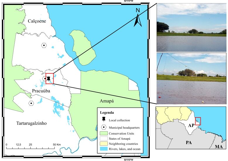
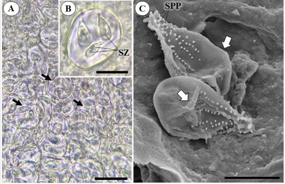
The tucunaré (Cichla sp.) is an Amazonian fish that is heavily commercialized in the state of Amapá, and it can be infected by a variety of parasites, including coccidia of the genus Calyptospora, which are identified at the genus level by analyzing the structures that comprise its morphology. This study aimed to describe the morphology and histopathology of Calyptospora sp. parasitism in Cichla monoculus Spix, 1929 in the Municipality of Pracuúba, Amapá, Brazil. Nine specimens were acquired from the Lake Sacaizal by artisanal fishermen and transported in isothermal boxes to the Integrated Morpho-molecular and Technologies Laboratory (LIMT) of the Federal Rural University of the Amazon in Belém, Pará, where they were necropsied. Fragments of the liver were removed to visualize cysts using light microscopy and processed for scanning electron microscopy and histology analyses. The analysis revealed that 66.6% of the fish examined had clusters of oocysts in the hepatic region, resulting in the formation of melanomacrophagic centers. The oocysts were sphere-like, with a diameter of 21 µm. They contained four pyriform sporocysts, 8.7 µm long and 4.9 µm wide, with sporopods in the posterior region.
期刊介绍:
La revista es un órgano de difusión del Colegio Brasileño de Parasitología Veterinaria, con una especificidad dentro de esa área, la difusión de los resultados de la investigación brasileña en las áreas de Helmintología, Protozoología, Entomología y agentes transmitidos por artrópodos, relacionados con la salud animal.


 分享
分享
 求助内容:
求助内容: 应助结果提醒方式:
 扫码关注我们
