Elizabeth G. Dunne MD , Cameron N. Fick MD , Brooke Mastrogiacomo MS , Kay See Tan PhD , Nicolas Toumbacaris MSPH , Stijn Vanstraelen MD , Gaetano Rocco MD , Jaime E. Chaft MD , Puneeth Iyengar MD , Daniel Gomez MD , Prasad S. Adusumilli MD , Bernard J. Park MD , James M. Isbell MD , Matthew J. Bott MD , Smita Sihag MD , Daniela Molena MD , James Huang MD , David R. Jones MD
{"title":"Clinicopathologic and genomic features associated with brain metastasis after resection of lung adenocarcinoma","authors":"Elizabeth G. Dunne MD , Cameron N. Fick MD , Brooke Mastrogiacomo MS , Kay See Tan PhD , Nicolas Toumbacaris MSPH , Stijn Vanstraelen MD , Gaetano Rocco MD , Jaime E. Chaft MD , Puneeth Iyengar MD , Daniel Gomez MD , Prasad S. Adusumilli MD , Bernard J. Park MD , James M. Isbell MD , Matthew J. Bott MD , Smita Sihag MD , Daniela Molena MD , James Huang MD , David R. Jones MD","doi":"10.1016/j.xjon.2024.09.030","DOIUrl":null,"url":null,"abstract":"<div><h3>Objective</h3><div>To identify clinicopathologic and genomic features associated with brain metastasis after resection of lung adenocarcinoma (LUAD) and to evaluate survival after brain metastasis.</div></div><div><h3>Methods</h3><div>Patients who underwent complete resection of stage I-IIIA LUAD between 2011 and 2020 were included. A subset of patients had broad-based panel next-generation sequencing performed on their tumors. Fine-Gray models for the development of brain metastasis were constructed, with death without brain metastasis as a competing risk.</div></div><div><h3>Results</h3><div>A total of 2660 patients were included. The median duration of follow-up was 71 months (95% confidence interval [CI], 69-73 months). The cumulative incidence of brain metastasis at 10 years was 9.8%. Among patients who developed a brain metastasis, the median time from surgery to brain metastasis was 21 months (interquartile range, 10-42 months). Higher maximum standardized uptake value of the primary tumor, neoadjuvant therapy, lymphovascular invasion, and stage III disease were associated with the development of brain metastasis. Among patients who underwent next-generation sequencing, a multivariable analysis identified neoadjuvant therapy, pathologic stage, and <em>TP53</em> mutations as associated with development of brain metastasis. The median survival after brain metastasis was 18 months (95% CI, 13-24 months). Better performance status, lack of extracranial metastasis, stereotactic radiosurgery, and targeted therapy were associated with better survival after brain metastasis.</div></div><div><h3>Conclusions</h3><div>Brain metastasis is common after complete resection of LUAD and often occurs within 2 years. Markers of aggressive tumor biology, including higher maximum standardized uptake value, lymphovascular invasion, and <em>TP53</em> mutations, and neoadjuvant therapy are associated with brain metastasis.</div></div>","PeriodicalId":74032,"journal":{"name":"JTCVS open","volume":"22 ","pages":"Pages 458-469"},"PeriodicalIF":1.9000,"publicationDate":"2024-12-01","publicationTypes":"Journal Article","fieldsOfStudy":null,"isOpenAccess":false,"openAccessPdf":"https://www.ncbi.nlm.nih.gov/pmc/articles/PMC11704575/pdf/","citationCount":"0","resultStr":null,"platform":"Semanticscholar","paperid":null,"PeriodicalName":"JTCVS open","FirstCategoryId":"1085","ListUrlMain":"https://www.sciencedirect.com/science/article/pii/S2666273624003504","RegionNum":0,"RegionCategory":null,"ArticlePicture":[],"TitleCN":null,"AbstractTextCN":null,"PMCID":null,"EPubDate":"2024/10/18 0:00:00","PubModel":"Epub","JCR":"","JCRName":"","Score":null,"Total":0}
引用次数: 0
Abstract
Objective
To identify clinicopathologic and genomic features associated with brain metastasis after resection of lung adenocarcinoma (LUAD) and to evaluate survival after brain metastasis.
Methods
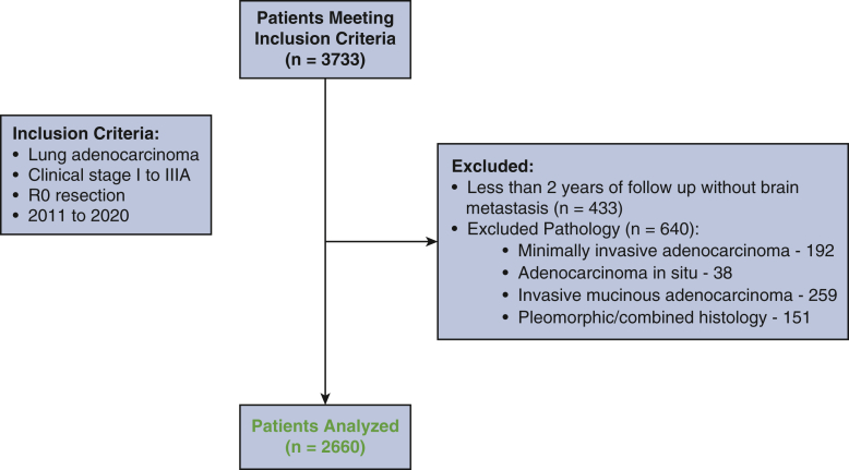
Patients who underwent complete resection of stage I-IIIA LUAD between 2011 and 2020 were included. A subset of patients had broad-based panel next-generation sequencing performed on their tumors. Fine-Gray models for the development of brain metastasis were constructed, with death without brain metastasis as a competing risk.
Results
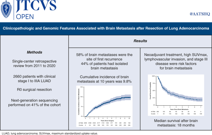
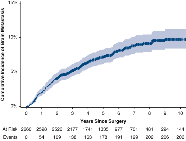
A total of 2660 patients were included. The median duration of follow-up was 71 months (95% confidence interval [CI], 69-73 months). The cumulative incidence of brain metastasis at 10 years was 9.8%. Among patients who developed a brain metastasis, the median time from surgery to brain metastasis was 21 months (interquartile range, 10-42 months). Higher maximum standardized uptake value of the primary tumor, neoadjuvant therapy, lymphovascular invasion, and stage III disease were associated with the development of brain metastasis. Among patients who underwent next-generation sequencing, a multivariable analysis identified neoadjuvant therapy, pathologic stage, and TP53 mutations as associated with development of brain metastasis. The median survival after brain metastasis was 18 months (95% CI, 13-24 months). Better performance status, lack of extracranial metastasis, stereotactic radiosurgery, and targeted therapy were associated with better survival after brain metastasis.
Conclusions
Brain metastasis is common after complete resection of LUAD and often occurs within 2 years. Markers of aggressive tumor biology, including higher maximum standardized uptake value, lymphovascular invasion, and TP53 mutations, and neoadjuvant therapy are associated with brain metastasis.


 分享
分享

 求助内容:
求助内容: 应助结果提醒方式:
 扫码关注我们
