Automatic segmentation model and machine learning model grounded in ultrasound radiomics for distinguishing between low malignant risk and intermediate-high malignant risk of adnexal masses.
{"title":"Automatic segmentation model and machine learning model grounded in ultrasound radiomics for distinguishing between low malignant risk and intermediate-high malignant risk of adnexal masses.","authors":"Lu Liu, Wenjun Cai, Feibo Zheng, Hongyan Tian, Yanping Li, Ting Wang, Xiaonan Chen, Wenjing Zhu","doi":"10.1186/s13244-024-01874-7","DOIUrl":null,"url":null,"abstract":"<p><strong>Objective: </strong>To develop an automatic segmentation model to delineate the adnexal masses and construct a machine learning model to differentiate between low malignant risk and intermediate-high malignant risk of adnexal masses based on ovarian-adnexal reporting and data system (O-RADS).</p><p><strong>Methods: </strong>A total of 663 ultrasound images of adnexal mass were collected and divided into two sets according to experienced radiologists: a low malignant risk set (n = 446) and an intermediate-high malignant risk set (n = 217). Deep learning segmentation models were trained and selected to automatically segment adnexal masses. Radiomics features were extracted utilizing a feature analysis system in Pyradiomics. Feature selection was conducted using the Spearman correlation analysis, Mann-Whitney U-test, and least absolute shrinkage and selection operator (LASSO) regression. A nomogram integrating radiomic and clinical features using a machine learning model was established and evaluated. The SHapley Additive exPlanations were used for model interpretability and visualization.</p><p><strong>Results: </strong>The FCN ResNet101 demonstrated the highest segmentation performance for adnexal masses (Dice similarity coefficient: 89.1%). Support vector machine achieved the best AUC (0.961, 95% CI: 0.925-0.996). The nomogram using the LightGBM algorithm reached the best AUC (0.966, 95% CI: 0.927-1.000). The diagnostic performance of the nomogram was comparable to that of experienced radiologists (p > 0.05) and outperformed that of less-experienced radiologists (p < 0.05). The model significantly improved the diagnostic accuracy of less-experienced radiologists.</p><p><strong>Conclusions: </strong>The segmentation model serves as a valuable tool for the automated delineation of adnexal lesions. The machine learning model exhibited commendable classification capability and outperformed the diagnostic performance of less-experienced radiologists.</p><p><strong>Critical relevance statement: </strong>The ultrasound radiomics-based machine learning model holds the potential to elevate the professional ability of less-experienced radiologists and can be used to assist in the clinical screening of ovarian cancer.</p><p><strong>Key points: </strong>We developed an image segmentation model to automatically delineate adnexal masses. We developed a model to classify adnexal masses based on O-RADS. The machine learning model has achieved commendable classification performance. The machine learning model possesses the capability to enhance the proficiency of less-experienced radiologists. We used SHapley Additive exPlanations to interpret and visualize the model.</p>","PeriodicalId":13639,"journal":{"name":"Insights into Imaging","volume":"16 1","pages":"14"},"PeriodicalIF":4.5000,"publicationDate":"2025-01-13","publicationTypes":"Journal Article","fieldsOfStudy":null,"isOpenAccess":false,"openAccessPdf":"https://www.ncbi.nlm.nih.gov/pmc/articles/PMC11729609/pdf/","citationCount":"0","resultStr":null,"platform":"Semanticscholar","paperid":null,"PeriodicalName":"Insights into Imaging","FirstCategoryId":"3","ListUrlMain":"https://doi.org/10.1186/s13244-024-01874-7","RegionNum":2,"RegionCategory":"医学","ArticlePicture":[],"TitleCN":null,"AbstractTextCN":null,"PMCID":null,"EPubDate":"","PubModel":"","JCR":"Q1","JCRName":"RADIOLOGY, NUCLEAR MEDICINE & MEDICAL IMAGING","Score":null,"Total":0}
引用次数: 0
Abstract
Objective: To develop an automatic segmentation model to delineate the adnexal masses and construct a machine learning model to differentiate between low malignant risk and intermediate-high malignant risk of adnexal masses based on ovarian-adnexal reporting and data system (O-RADS).
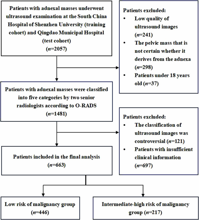
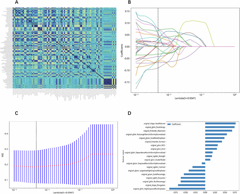
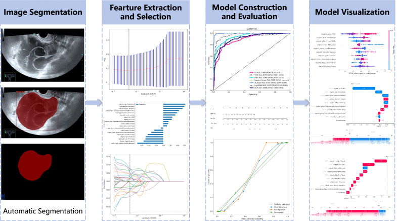
Methods: A total of 663 ultrasound images of adnexal mass were collected and divided into two sets according to experienced radiologists: a low malignant risk set (n = 446) and an intermediate-high malignant risk set (n = 217). Deep learning segmentation models were trained and selected to automatically segment adnexal masses. Radiomics features were extracted utilizing a feature analysis system in Pyradiomics. Feature selection was conducted using the Spearman correlation analysis, Mann-Whitney U-test, and least absolute shrinkage and selection operator (LASSO) regression. A nomogram integrating radiomic and clinical features using a machine learning model was established and evaluated. The SHapley Additive exPlanations were used for model interpretability and visualization.
Results: The FCN ResNet101 demonstrated the highest segmentation performance for adnexal masses (Dice similarity coefficient: 89.1%). Support vector machine achieved the best AUC (0.961, 95% CI: 0.925-0.996). The nomogram using the LightGBM algorithm reached the best AUC (0.966, 95% CI: 0.927-1.000). The diagnostic performance of the nomogram was comparable to that of experienced radiologists (p > 0.05) and outperformed that of less-experienced radiologists (p < 0.05). The model significantly improved the diagnostic accuracy of less-experienced radiologists.
Conclusions: The segmentation model serves as a valuable tool for the automated delineation of adnexal lesions. The machine learning model exhibited commendable classification capability and outperformed the diagnostic performance of less-experienced radiologists.
Critical relevance statement: The ultrasound radiomics-based machine learning model holds the potential to elevate the professional ability of less-experienced radiologists and can be used to assist in the clinical screening of ovarian cancer.
Key points: We developed an image segmentation model to automatically delineate adnexal masses. We developed a model to classify adnexal masses based on O-RADS. The machine learning model has achieved commendable classification performance. The machine learning model possesses the capability to enhance the proficiency of less-experienced radiologists. We used SHapley Additive exPlanations to interpret and visualize the model.
期刊介绍:
Insights into Imaging (I³) is a peer-reviewed open access journal published under the brand SpringerOpen. All content published in the journal is freely available online to anyone, anywhere!
I³ continuously updates scientific knowledge and progress in best-practice standards in radiology through the publication of original articles and state-of-the-art reviews and opinions, along with recommendations and statements from the leading radiological societies in Europe.
Founded by the European Society of Radiology (ESR), I³ creates a platform for educational material, guidelines and recommendations, and a forum for topics of controversy.
A balanced combination of review articles, original papers, short communications from European radiological congresses and information on society matters makes I³ an indispensable source for current information in this field.
I³ is owned by the ESR, however authors retain copyright to their article according to the Creative Commons Attribution License (see Copyright and License Agreement). All articles can be read, redistributed and reused for free, as long as the author of the original work is cited properly.
The open access fees (article-processing charges) for this journal are kindly sponsored by ESR for all Members.
The journal went open access in 2012, which means that all articles published since then are freely available online.


 分享
分享
 求助内容:
求助内容: 应助结果提醒方式:
 扫码关注我们
