{"title":"A Rare Case of the Ipsilateral Paraspinal Muscle Abscess Communicating with a Psoas Major Abscess: A Case Report.","authors":"Junya Kusakabe, Katsushi Suzuki, Masami Hosaka","doi":"10.13107/jocr.2025.v15.i01.5158","DOIUrl":null,"url":null,"abstract":"<p><strong>Background: </strong>Paraspinal muscle abscesses are rare, and generally occur due to injections or hematogenous dissemination. Here, we describe a rare case of a paraspinal muscle and the ipsilateral psoas major abscess in the lumbar region that communicated via the interspaces of the costal processes.</p><p><strong>Case report: </strong>An 83-year-old man with poorly controlled diabetes mellitus and no history of puncture complained of right low back pain for the past 2 months. He was diagnosed with pyelonephritis and referred to our department for close examination of the low back pain. Magnetic resonance imaging revealed a paraspinal muscle and an ipsilateral psoas major abscess in the lumbar region, which communicated through the interspaces of the costal processes. A definitive diagnosis was made using percutaneous aspiration and the patient was successfully treated conservatively.</p><p><strong>Conclusion: </strong>This case is very rare and impressive because the paraspinal muscle abscess directly communicated with the ipsilateral psoas major muscle abscess. Spinal infections should always be considered in the differential diagnosis of low back pain, particularly in the absence of long-term improvement. Local physical examinations are essential when examining patients with low back pain. Conservative treatment is effective, even if the abscess is extensive.</p>","PeriodicalId":16647,"journal":{"name":"Journal of Orthopaedic Case Reports","volume":"15 1","pages":"150-154"},"PeriodicalIF":0.0000,"publicationDate":"2025-01-01","publicationTypes":"Journal Article","fieldsOfStudy":null,"isOpenAccess":false,"openAccessPdf":"https://www.ncbi.nlm.nih.gov/pmc/articles/PMC11723724/pdf/","citationCount":"0","resultStr":null,"platform":"Semanticscholar","paperid":null,"PeriodicalName":"Journal of Orthopaedic Case Reports","FirstCategoryId":"1085","ListUrlMain":"https://doi.org/10.13107/jocr.2025.v15.i01.5158","RegionNum":0,"RegionCategory":null,"ArticlePicture":[],"TitleCN":null,"AbstractTextCN":null,"PMCID":null,"EPubDate":"","PubModel":"","JCR":"","JCRName":"","Score":null,"Total":0}
引用次数: 0
Abstract
Background: Paraspinal muscle abscesses are rare, and generally occur due to injections or hematogenous dissemination. Here, we describe a rare case of a paraspinal muscle and the ipsilateral psoas major abscess in the lumbar region that communicated via the interspaces of the costal processes.
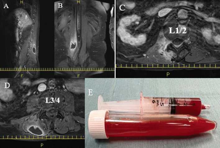
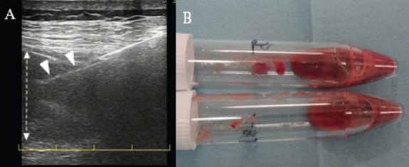
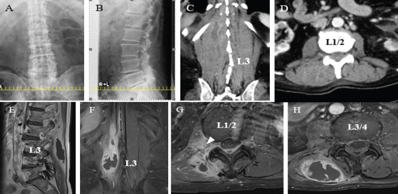
Case report: An 83-year-old man with poorly controlled diabetes mellitus and no history of puncture complained of right low back pain for the past 2 months. He was diagnosed with pyelonephritis and referred to our department for close examination of the low back pain. Magnetic resonance imaging revealed a paraspinal muscle and an ipsilateral psoas major abscess in the lumbar region, which communicated through the interspaces of the costal processes. A definitive diagnosis was made using percutaneous aspiration and the patient was successfully treated conservatively.
Conclusion: This case is very rare and impressive because the paraspinal muscle abscess directly communicated with the ipsilateral psoas major muscle abscess. Spinal infections should always be considered in the differential diagnosis of low back pain, particularly in the absence of long-term improvement. Local physical examinations are essential when examining patients with low back pain. Conservative treatment is effective, even if the abscess is extensive.


 分享
分享

 求助内容:
求助内容: 应助结果提醒方式:
 扫码关注我们
