{"title":"Seminal vesicle schwannoma with chronic hemorrhage.","authors":"Ting Li, Yong-Fang Zhang, Zhou-Liang Yang, Zhuo-Peng Ying","doi":"10.17712/nsj.2025.1.20240073","DOIUrl":null,"url":null,"abstract":"<p><p>Schwannomas are benign tumors originating from Schwann cells, with seminal vesicle schwannomas being exceedingly rare. This report describes a 54-year-old man with an incidental discovery of a right-sided seminal vesicle mass during a routine ultrasound examination. Further imaging, including MRI and contrast-enhanced CT scans, revealed a well-defined, encapsulated mass with heterogeneous signal intensity suggestive of schwannoma. Histopathological examination confirmed schwannoma, marked by positive S-100 and NSE expression and a Ki67 index of 5%. The patient underwent successful laparoscopic resection without complications and remained asymptomatic for over a month. This case underscores the importance of integrating MRI and histopathological findings to accurately diagnose seminal vesicle schwannomas and guide appropriate surgical management, highlighting the need for increased clinician awareness of this rare tumor.</p>","PeriodicalId":19284,"journal":{"name":"Neurosciences","volume":"30 1","pages":"59-63"},"PeriodicalIF":1.3000,"publicationDate":"2025-01-01","publicationTypes":"Journal Article","fieldsOfStudy":null,"isOpenAccess":false,"openAccessPdf":"https://www.ncbi.nlm.nih.gov/pmc/articles/PMC11753593/pdf/","citationCount":"0","resultStr":null,"platform":"Semanticscholar","paperid":null,"PeriodicalName":"Neurosciences","FirstCategoryId":"3","ListUrlMain":"https://doi.org/10.17712/nsj.2025.1.20240073","RegionNum":4,"RegionCategory":"医学","ArticlePicture":[],"TitleCN":null,"AbstractTextCN":null,"PMCID":null,"EPubDate":"","PubModel":"","JCR":"Q4","JCRName":"CLINICAL NEUROLOGY","Score":null,"Total":0}
引用次数: 0
Abstract
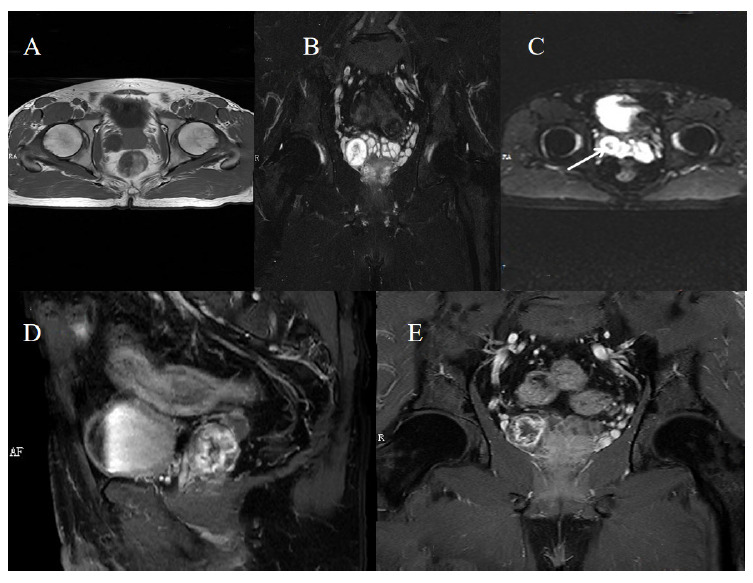
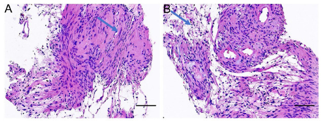
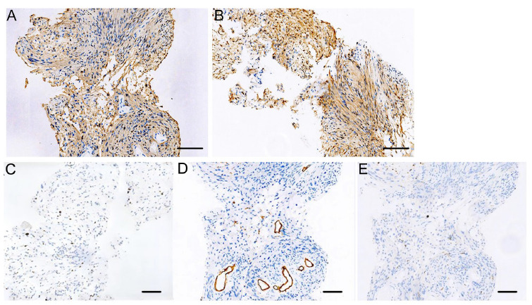
Schwannomas are benign tumors originating from Schwann cells, with seminal vesicle schwannomas being exceedingly rare. This report describes a 54-year-old man with an incidental discovery of a right-sided seminal vesicle mass during a routine ultrasound examination. Further imaging, including MRI and contrast-enhanced CT scans, revealed a well-defined, encapsulated mass with heterogeneous signal intensity suggestive of schwannoma. Histopathological examination confirmed schwannoma, marked by positive S-100 and NSE expression and a Ki67 index of 5%. The patient underwent successful laparoscopic resection without complications and remained asymptomatic for over a month. This case underscores the importance of integrating MRI and histopathological findings to accurately diagnose seminal vesicle schwannomas and guide appropriate surgical management, highlighting the need for increased clinician awareness of this rare tumor.
期刊介绍:
Neurosciences is an open access, peer-reviewed, quarterly publication. Authors are invited to submit for publication articles reporting original work related to the nervous system, e.g., neurology, neurophysiology, neuroradiology, neurosurgery, neurorehabilitation, neurooncology, neuropsychiatry, and neurogenetics, etc. Basic research withclear clinical implications will also be considered. Review articles of current interest and high standard are welcomed for consideration. Prospective workshould not be backdated. There are also sections for Case Reports, Brief Communication, Correspondence, and medical news items. To promote continuous education, training, and learning, we include Clinical Images and MCQ’s. Highlights of international and regional meetings of interest, and specialized supplements will also be considered. All submissions must conform to the Uniform Requirements.


 分享
分享
 求助内容:
求助内容: 应助结果提醒方式:
 扫码关注我们
