{"title":"Imaging of Venolymphatic Malformation in a Child Extending from Inguinoscrotal Region to Thoracic Region: Case Report with Review of Literature.","authors":"Mohammad Ameen Abdus Salam Ansari, Avinash Parshuram Dhok, Pooja Giridhar Ladke, Nitin Shinde","doi":"10.4103/jmu.jmu_44_23","DOIUrl":null,"url":null,"abstract":"<p><p>Vascular malformations (VM) are structural malformations of vascular development causing soft-tissue abnormality with functional and esthetic impairment. They are named by their predominant vessel type as arterial, venous, lymphatic, or mixed types. VM extending from the inguinoscrotal to the thoracic region are extremely rare presentation. We present a rare case of veno-lymphatic malformation in the inguinoscrotal region, which is extending superiorly up to the right thorax in a 14-year-old male child who presented with a large swelling in the bilateral inguinoscrotal region and reddish-brown colored skin patches over the right anterior and lateral thoracoabdominal region. The diagnosis was suggested by ultrasonography and confirmed by computed tomography and magnetic resonance imaging.</p>","PeriodicalId":45466,"journal":{"name":"Journal of Medical Ultrasound","volume":"32 4","pages":"345-347"},"PeriodicalIF":0.8000,"publicationDate":"2023-10-06","publicationTypes":"Journal Article","fieldsOfStudy":null,"isOpenAccess":false,"openAccessPdf":"https://www.ncbi.nlm.nih.gov/pmc/articles/PMC11717088/pdf/","citationCount":"0","resultStr":null,"platform":"Semanticscholar","paperid":null,"PeriodicalName":"Journal of Medical Ultrasound","FirstCategoryId":"1085","ListUrlMain":"https://doi.org/10.4103/jmu.jmu_44_23","RegionNum":0,"RegionCategory":null,"ArticlePicture":[],"TitleCN":null,"AbstractTextCN":null,"PMCID":null,"EPubDate":"2024/10/1 0:00:00","PubModel":"eCollection","JCR":"Q4","JCRName":"RADIOLOGY, NUCLEAR MEDICINE & MEDICAL IMAGING","Score":null,"Total":0}
引用次数: 0
Abstract
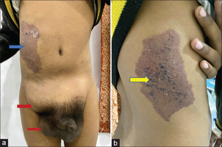
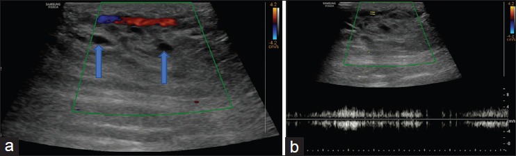
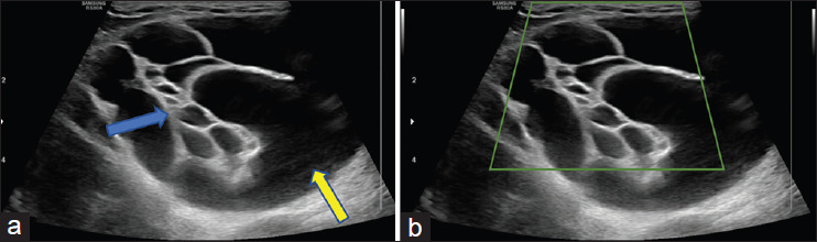
Vascular malformations (VM) are structural malformations of vascular development causing soft-tissue abnormality with functional and esthetic impairment. They are named by their predominant vessel type as arterial, venous, lymphatic, or mixed types. VM extending from the inguinoscrotal to the thoracic region are extremely rare presentation. We present a rare case of veno-lymphatic malformation in the inguinoscrotal region, which is extending superiorly up to the right thorax in a 14-year-old male child who presented with a large swelling in the bilateral inguinoscrotal region and reddish-brown colored skin patches over the right anterior and lateral thoracoabdominal region. The diagnosis was suggested by ultrasonography and confirmed by computed tomography and magnetic resonance imaging.
期刊介绍:
The Journal of Medical Ultrasound is the peer-reviewed publication of the Asian Federation of Societies for Ultrasound in Medicine and Biology, and the Chinese Taipei Society of Ultrasound in Medicine. Its aim is to promote clinical and scientific research in ultrasonography, and to serve as a channel of communication among sonologists, sonographers, and medical ultrasound physicians in the Asia-Pacific region and wider international community. The Journal invites original contributions relating to the clinical and laboratory investigations and applications of ultrasonography.


 分享
分享
 求助内容:
求助内容: 应助结果提醒方式:
 扫码关注我们
