Relationship between the location of the popliteal artery and the tibial osteotomy plane in patients with medial and lateral unicompartmental knee arthroplasty: A retrospective analysis of preoperative magnetic resonance imaging and intraoperative findings.
{"title":"Relationship between the location of the popliteal artery and the tibial osteotomy plane in patients with medial and lateral unicompartmental knee arthroplasty: A retrospective analysis of preoperative magnetic resonance imaging and intraoperative findings.","authors":"Tatsuya Kubo, Tsuneari Takahashi, Yuya Kimura, Takashi Ajiki, Eri Yasuda, Katsushi Takeshita","doi":"10.1051/sicotj/2024058","DOIUrl":null,"url":null,"abstract":"<p><strong>Purpose: </strong>To clarify the location of the popliteal artery (PA) is relative to the tibial osteotomy plane in patients with medial and lateral unicompartmental knee osteoarthritis (KOA) undergoing UKA.</p><p><strong>Methods: </strong>Preoperative MRI and postoperative radiographs obtained from 50 patients with unicompartmental KOA who underwent fixed-bearing UKA were analyzed. The amount of tibial resection was determined from the surgical records, and a line was drawn parallel to the tibial posterior tilt angle on the sagittal MR image to create a virtual tibial cut line. The tibial resection width measured from the anteroposterior image of the postoperative radiograph was projected onto the transverse plane containing the intersection between the virtual tibial cut line and the posterior tibial cortex, after which a line was drawn parallel to the medial or lateral intercondylar ridge. We then determined whether the PA was within an extension of the osteotomy area. The shortest distance (Distance 1) between the posterior tibial cortex and the PA within the osteotomy area was measured. In addition, the shortest distance between the line extending the osteotomy posteriorly and the PA was measured (Distance 2).</p><p><strong>Results: </strong>The medial UKA (group M) and lateral UKA (group L) group comprised 41 and 9 cases. The percentage of PA located behind the osteotomy plane was significantly higher in group L than in group M [6/9 knees (66.7%) vs. 2/41 knees (4.9%); P < 0.001]. The distance 1 was 12.6 (4.3) mm in group M and 7.9 (3.7) mm in group L (P = 0.004). The distance2 was 11.1 (4.9) mm in group M and 2.6 (3.5) mm in group L (P < 0.001).</p><p><strong>Conclusion: </strong>During lateral UKA, the PA was often located behind the tibial osteotomy plane and close to the posterior tibial cortex. Nearly 5% of medial UKAs, the artery was located behind the osteotomy plane.</p><p><strong>Level of evidence: </strong>Retrospective comparative LEVEL III study.</p>","PeriodicalId":46378,"journal":{"name":"SICOT-J","volume":"11 ","pages":"1"},"PeriodicalIF":2.3000,"publicationDate":"2025-01-01","publicationTypes":"Journal Article","fieldsOfStudy":null,"isOpenAccess":false,"openAccessPdf":"https://www.ncbi.nlm.nih.gov/pmc/articles/PMC11727081/pdf/","citationCount":"0","resultStr":null,"platform":"Semanticscholar","paperid":null,"PeriodicalName":"SICOT-J","FirstCategoryId":"1085","ListUrlMain":"https://doi.org/10.1051/sicotj/2024058","RegionNum":0,"RegionCategory":null,"ArticlePicture":[],"TitleCN":null,"AbstractTextCN":null,"PMCID":null,"EPubDate":"2025/1/13 0:00:00","PubModel":"Epub","JCR":"Q2","JCRName":"ORTHOPEDICS","Score":null,"Total":0}
引用次数: 0
Abstract
Purpose: To clarify the location of the popliteal artery (PA) is relative to the tibial osteotomy plane in patients with medial and lateral unicompartmental knee osteoarthritis (KOA) undergoing UKA.
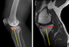
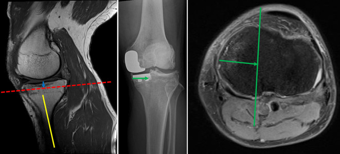
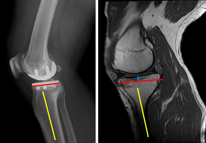
Methods: Preoperative MRI and postoperative radiographs obtained from 50 patients with unicompartmental KOA who underwent fixed-bearing UKA were analyzed. The amount of tibial resection was determined from the surgical records, and a line was drawn parallel to the tibial posterior tilt angle on the sagittal MR image to create a virtual tibial cut line. The tibial resection width measured from the anteroposterior image of the postoperative radiograph was projected onto the transverse plane containing the intersection between the virtual tibial cut line and the posterior tibial cortex, after which a line was drawn parallel to the medial or lateral intercondylar ridge. We then determined whether the PA was within an extension of the osteotomy area. The shortest distance (Distance 1) between the posterior tibial cortex and the PA within the osteotomy area was measured. In addition, the shortest distance between the line extending the osteotomy posteriorly and the PA was measured (Distance 2).
Results: The medial UKA (group M) and lateral UKA (group L) group comprised 41 and 9 cases. The percentage of PA located behind the osteotomy plane was significantly higher in group L than in group M [6/9 knees (66.7%) vs. 2/41 knees (4.9%); P < 0.001]. The distance 1 was 12.6 (4.3) mm in group M and 7.9 (3.7) mm in group L (P = 0.004). The distance2 was 11.1 (4.9) mm in group M and 2.6 (3.5) mm in group L (P < 0.001).
Conclusion: During lateral UKA, the PA was often located behind the tibial osteotomy plane and close to the posterior tibial cortex. Nearly 5% of medial UKAs, the artery was located behind the osteotomy plane.
Level of evidence: Retrospective comparative LEVEL III study.


 分享
分享
 求助内容:
求助内容: 应助结果提醒方式:
 扫码关注我们
