Junchao Ma, Enyu Yuan, Shijian Feng, Jin Yao, Chunlei He, Yuntian Chen, Bin Song
{"title":"Diagnostic performance of CT for extrarenal fat invasion in renal cell carcinoma: a meta-analysis and systematic review.","authors":"Junchao Ma, Enyu Yuan, Shijian Feng, Jin Yao, Chunlei He, Yuntian Chen, Bin Song","doi":"10.1186/s13244-024-01889-0","DOIUrl":null,"url":null,"abstract":"<p><strong>Objectives: </strong>Renal cell carcinoma (RCC) with extrarenal fat (perinephric or renal sinus fat) invasion is the main evidence for the T3a stage. Currently, computed tomography (CT) is still the primary modality for staging RCC. This study aims to determine the diagnostic performance of CT in RCC patients with extrarenal fat invasion.</p><p><strong>Methods: </strong>The PubMed, Web of Science, Cochrane Library, and EMBASE databases were systematically searched up to October 11, 2023. Study quality was assessed by the QUADAS-2 tool. Standard methods recommended for meta-analyses of diagnostic evaluation were used. Heterogeneity was analyzed through meta-regression analysis.</p><p><strong>Results: </strong>Fifteen studies were included in this meta-analysis. Among them, six studies focused on perinephric fat invasion (PFI) only, four on renal sinus fat invasion (RSFI) only, and five on both. Pooled weighted estimates of sensitivity, specificity, area of SROC curve, PLR, and negative likelihood ratio (NLR) of CT for PFI were 0.69 (95% CI: 0.55-0.79), 0.82 (95% CI: 0.69-0.90), 0.81 (95% CI: 0.77-0.84), 3.85 (95% CI: 2.22-6.67), and 0.38 (95% CI: 0.27-0.55). Pooled weighted estimates of sensitivity, specificity, area of SROC curve, PLR, and NLR of CT for RSFI were 0.81 (95% CI: 0.76-0.85), 0.79 (95% CI: 0.66-0.88), 0.82 (95% CI: 0.78-0.85), 3.91 (95% CI: 2.26-6.77), and 0.24 (95% CI: 0.18-0.31).</p><p><strong>Conclusion: </strong>CT has the ability to detect the PFI and RSFI in patients with RCC. However, the diagnostic performance of CT has suffered from the limitation of slightly lower accuracy, resulting from the low positive sample in the current studies. Additionally, the current PLR is low.</p><p><strong>Critical relevance statement: </strong>This study provides radiologists and urologists with a systematic and comprehensive summary of CT and CT-related morphological features in assessing extrarenal fat invasion in patients with RCC.</p><p><strong>Key points: </strong>CT can detect extrarenal fat invasion in patients with RCC, but the diagnostic performance is inconsistent. The diagnostic performance of CT is acceptable, but primarily affected by the low positive rate of included patients. Further large-scale trials are necessary to determine the true diagnostic capabilities of CT for extrarenal fat invasion.</p>","PeriodicalId":13639,"journal":{"name":"Insights into Imaging","volume":"16 1","pages":"19"},"PeriodicalIF":4.5000,"publicationDate":"2025-01-15","publicationTypes":"Journal Article","fieldsOfStudy":null,"isOpenAccess":false,"openAccessPdf":"https://www.ncbi.nlm.nih.gov/pmc/articles/PMC11735820/pdf/","citationCount":"0","resultStr":null,"platform":"Semanticscholar","paperid":null,"PeriodicalName":"Insights into Imaging","FirstCategoryId":"3","ListUrlMain":"https://doi.org/10.1186/s13244-024-01889-0","RegionNum":2,"RegionCategory":"医学","ArticlePicture":[],"TitleCN":null,"AbstractTextCN":null,"PMCID":null,"EPubDate":"","PubModel":"","JCR":"Q1","JCRName":"RADIOLOGY, NUCLEAR MEDICINE & MEDICAL IMAGING","Score":null,"Total":0}
引用次数: 0
Abstract
Objectives: Renal cell carcinoma (RCC) with extrarenal fat (perinephric or renal sinus fat) invasion is the main evidence for the T3a stage. Currently, computed tomography (CT) is still the primary modality for staging RCC. This study aims to determine the diagnostic performance of CT in RCC patients with extrarenal fat invasion.
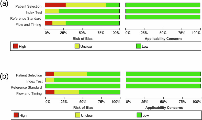
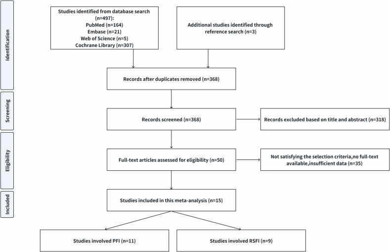
Methods: The PubMed, Web of Science, Cochrane Library, and EMBASE databases were systematically searched up to October 11, 2023. Study quality was assessed by the QUADAS-2 tool. Standard methods recommended for meta-analyses of diagnostic evaluation were used. Heterogeneity was analyzed through meta-regression analysis.
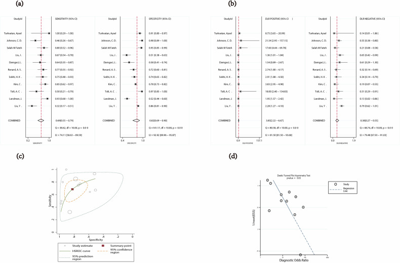
Results: Fifteen studies were included in this meta-analysis. Among them, six studies focused on perinephric fat invasion (PFI) only, four on renal sinus fat invasion (RSFI) only, and five on both. Pooled weighted estimates of sensitivity, specificity, area of SROC curve, PLR, and negative likelihood ratio (NLR) of CT for PFI were 0.69 (95% CI: 0.55-0.79), 0.82 (95% CI: 0.69-0.90), 0.81 (95% CI: 0.77-0.84), 3.85 (95% CI: 2.22-6.67), and 0.38 (95% CI: 0.27-0.55). Pooled weighted estimates of sensitivity, specificity, area of SROC curve, PLR, and NLR of CT for RSFI were 0.81 (95% CI: 0.76-0.85), 0.79 (95% CI: 0.66-0.88), 0.82 (95% CI: 0.78-0.85), 3.91 (95% CI: 2.26-6.77), and 0.24 (95% CI: 0.18-0.31).
Conclusion: CT has the ability to detect the PFI and RSFI in patients with RCC. However, the diagnostic performance of CT has suffered from the limitation of slightly lower accuracy, resulting from the low positive sample in the current studies. Additionally, the current PLR is low.
Critical relevance statement: This study provides radiologists and urologists with a systematic and comprehensive summary of CT and CT-related morphological features in assessing extrarenal fat invasion in patients with RCC.
Key points: CT can detect extrarenal fat invasion in patients with RCC, but the diagnostic performance is inconsistent. The diagnostic performance of CT is acceptable, but primarily affected by the low positive rate of included patients. Further large-scale trials are necessary to determine the true diagnostic capabilities of CT for extrarenal fat invasion.
期刊介绍:
Insights into Imaging (I³) is a peer-reviewed open access journal published under the brand SpringerOpen. All content published in the journal is freely available online to anyone, anywhere!
I³ continuously updates scientific knowledge and progress in best-practice standards in radiology through the publication of original articles and state-of-the-art reviews and opinions, along with recommendations and statements from the leading radiological societies in Europe.
Founded by the European Society of Radiology (ESR), I³ creates a platform for educational material, guidelines and recommendations, and a forum for topics of controversy.
A balanced combination of review articles, original papers, short communications from European radiological congresses and information on society matters makes I³ an indispensable source for current information in this field.
I³ is owned by the ESR, however authors retain copyright to their article according to the Creative Commons Attribution License (see Copyright and License Agreement). All articles can be read, redistributed and reused for free, as long as the author of the original work is cited properly.
The open access fees (article-processing charges) for this journal are kindly sponsored by ESR for all Members.
The journal went open access in 2012, which means that all articles published since then are freely available online.


 分享
分享
 求助内容:
求助内容: 应助结果提醒方式:
 扫码关注我们
