{"title":"Influence of Axial Rotation Between the Femoral Neck and Ankle Joint on Kinematics in Normal Knees: A Cross-Sectional Study.","authors":"Kenichi Kono, Shuji Taketomi, Takaharu Yamazaki, Tomofumi Kage, Masashi Tamaki, Hiroshi Inui, Sakae Tanaka, Tetsuya Tomita","doi":"10.5435/JAAOSGlobal-D-24-00169","DOIUrl":null,"url":null,"abstract":"<p><strong>Background: </strong>The effect of axial rotation between the femoral neck and ankle joint (total rotation [TR]) on normal knees is unknown. Therefore, this study aimed to investigate the TR effect on normal knee kinematics.</p><p><strong>Methods: </strong>Volunteers were divided into groups large (L), intermediate (I), and small (S), using hierarchical cluster analysis based on TR in the standing position. TR was measured using three-dimensional (3D) bone models generated from CT. A two-dimensional to 3-dimensional registration technique was used to assess the spatial position and femur and tibia orientation during squat. The axial rotation, varus-valgus alignment, and anterior-posterior translation of the femur relative to the tibia were evaluated.</p><p><strong>Results: </strong>Group L had the highest TR, whereas group S had the lowest TR (L: 36.6° ± 6.0°, I: 23.2° ± 3.0°, and S: 13.8° ± 5.1°). Above 50° of flexion, femoral external rotation was greater in group S than in groups L and I. From 40° to 110°, the medial side was more anterior in group L than in groups I and S, whereas the lateral side was more posterior in group S than in groups L and I.</p><p><strong>Conclusions: </strong>Individuals with larger TR had more femur anterior-medial translation relative to the tibia.</p>","PeriodicalId":45062,"journal":{"name":"Journal of the American Academy of Orthopaedic Surgeons Global Research and Reviews","volume":"9 1","pages":""},"PeriodicalIF":2.1000,"publicationDate":"2025-01-07","publicationTypes":"Journal Article","fieldsOfStudy":null,"isOpenAccess":false,"openAccessPdf":"https://www.ncbi.nlm.nih.gov/pmc/articles/PMC11709167/pdf/","citationCount":"0","resultStr":null,"platform":"Semanticscholar","paperid":null,"PeriodicalName":"Journal of the American Academy of Orthopaedic Surgeons Global Research and Reviews","FirstCategoryId":"1085","ListUrlMain":"https://doi.org/10.5435/JAAOSGlobal-D-24-00169","RegionNum":0,"RegionCategory":null,"ArticlePicture":[],"TitleCN":null,"AbstractTextCN":null,"PMCID":null,"EPubDate":"2025/1/1 0:00:00","PubModel":"eCollection","JCR":"Q2","JCRName":"ORTHOPEDICS","Score":null,"Total":0}
引用次数: 0
Abstract
Background: The effect of axial rotation between the femoral neck and ankle joint (total rotation [TR]) on normal knees is unknown. Therefore, this study aimed to investigate the TR effect on normal knee kinematics.
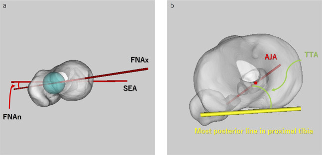
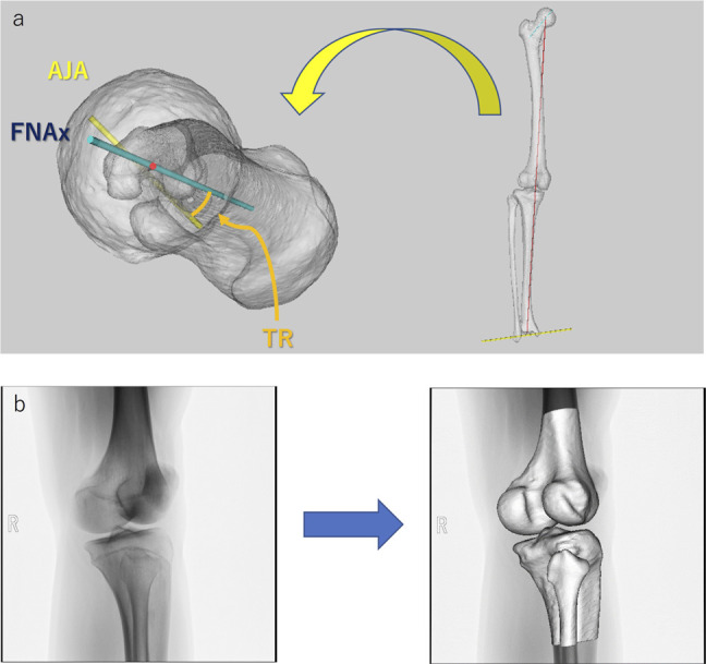
Methods: Volunteers were divided into groups large (L), intermediate (I), and small (S), using hierarchical cluster analysis based on TR in the standing position. TR was measured using three-dimensional (3D) bone models generated from CT. A two-dimensional to 3-dimensional registration technique was used to assess the spatial position and femur and tibia orientation during squat. The axial rotation, varus-valgus alignment, and anterior-posterior translation of the femur relative to the tibia were evaluated.
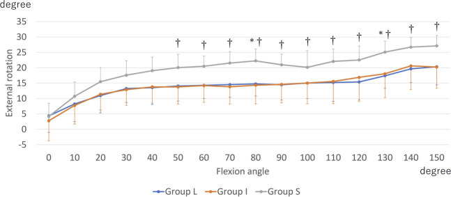
Results: Group L had the highest TR, whereas group S had the lowest TR (L: 36.6° ± 6.0°, I: 23.2° ± 3.0°, and S: 13.8° ± 5.1°). Above 50° of flexion, femoral external rotation was greater in group S than in groups L and I. From 40° to 110°, the medial side was more anterior in group L than in groups I and S, whereas the lateral side was more posterior in group S than in groups L and I.
Conclusions: Individuals with larger TR had more femur anterior-medial translation relative to the tibia.


 分享
分享

 求助内容:
求助内容: 应助结果提醒方式:
 扫码关注我们
