Abdul Veli Ismailoglu, Umit Suleyman Sehirli, Dilruba Ayingen, Alp Bayramoglu, Cemre Savasan, Baris Kocaoglu
{"title":"The Topographic Map of the Midfoot: Implication for Improving Safety of Dorsal Approach of Midfoot Surgeries.","authors":"Abdul Veli Ismailoglu, Umit Suleyman Sehirli, Dilruba Ayingen, Alp Bayramoglu, Cemre Savasan, Baris Kocaoglu","doi":"10.5435/JAAOSGlobal-D-24-00339","DOIUrl":null,"url":null,"abstract":"<p><strong>Purpose: </strong>The surgical approach for midfoot injuries classically requires dual dorsal incision and identification of the neurovascular structures that are susceptible to injury during the surgery. The aim of this study was to map the topographic anatomy of the dorsum of the foot along with tarsal joints for the dorsal approach of midfoot surgery that would facilitate the surgery and minimize the risk of neurovascular injuries for surgeons who specially focus on foot and ankle injuries.</p><p><strong>Methods: </strong>The dorsum of the foot was evaluated in 12 feet injected with latex containing a red colorant to visualize the arterial vessels. The navicular line, originating from the navicular tuberosity (NT) and passing over the dorsum of the foot, was used as a reference line. Dorsal foot neurovascular structures including cutaneous branches and muscles were mapped with respect to the tarsal joints and navicular line.</p><p><strong>Results: </strong>The deep peroneal nerve and dorsalis pedis artery were coursing between the base of the first and second metatarsal bones over which the tendon of the extensor hallucis brevis muscle was passing. The tendon of extensor hallucis brevis was crossing over the deep peroneal nerve and dorsalis pedis artery 55.2 mm superior and 45.0 mm lateral, respectively, from the NT.</p><p><strong>Conclusion: </strong>This cadaver study supplies a detailed topographic map of the dorsum of the foot using the tarsal joints and NT as landmarks for protecting the neurovascular structures to facilitate midfoot surgeries for sports medicine surgeons who specially focus on foot and ankle injuries.</p>","PeriodicalId":45062,"journal":{"name":"Journal of the American Academy of Orthopaedic Surgeons Global Research and Reviews","volume":"9 1","pages":""},"PeriodicalIF":2.1000,"publicationDate":"2025-01-07","publicationTypes":"Journal Article","fieldsOfStudy":null,"isOpenAccess":false,"openAccessPdf":"https://www.ncbi.nlm.nih.gov/pmc/articles/PMC11709205/pdf/","citationCount":"0","resultStr":null,"platform":"Semanticscholar","paperid":null,"PeriodicalName":"Journal of the American Academy of Orthopaedic Surgeons Global Research and Reviews","FirstCategoryId":"1085","ListUrlMain":"https://doi.org/10.5435/JAAOSGlobal-D-24-00339","RegionNum":0,"RegionCategory":null,"ArticlePicture":[],"TitleCN":null,"AbstractTextCN":null,"PMCID":null,"EPubDate":"2025/1/1 0:00:00","PubModel":"eCollection","JCR":"Q2","JCRName":"ORTHOPEDICS","Score":null,"Total":0}
引用次数: 0
Abstract
Purpose: The surgical approach for midfoot injuries classically requires dual dorsal incision and identification of the neurovascular structures that are susceptible to injury during the surgery. The aim of this study was to map the topographic anatomy of the dorsum of the foot along with tarsal joints for the dorsal approach of midfoot surgery that would facilitate the surgery and minimize the risk of neurovascular injuries for surgeons who specially focus on foot and ankle injuries.
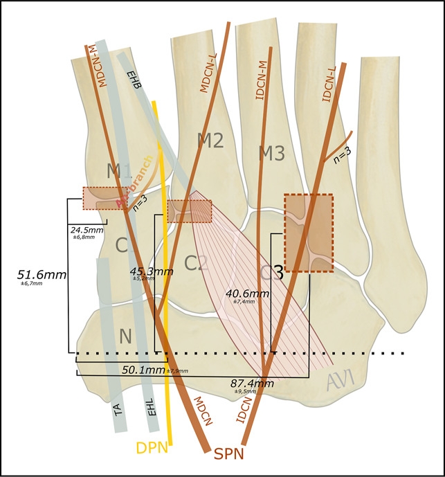
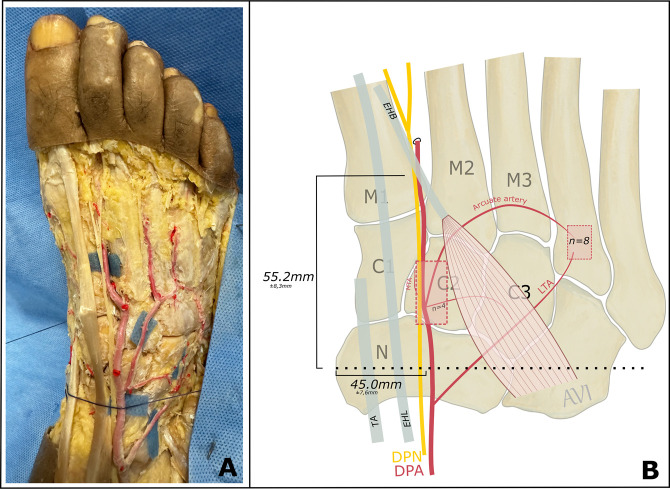
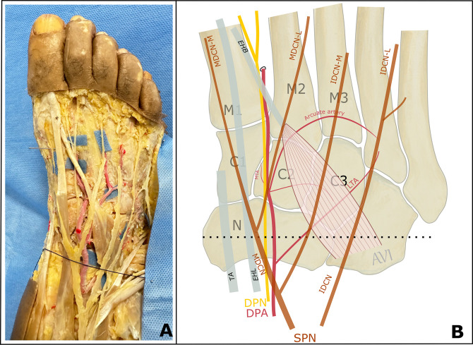
Methods: The dorsum of the foot was evaluated in 12 feet injected with latex containing a red colorant to visualize the arterial vessels. The navicular line, originating from the navicular tuberosity (NT) and passing over the dorsum of the foot, was used as a reference line. Dorsal foot neurovascular structures including cutaneous branches and muscles were mapped with respect to the tarsal joints and navicular line.
Results: The deep peroneal nerve and dorsalis pedis artery were coursing between the base of the first and second metatarsal bones over which the tendon of the extensor hallucis brevis muscle was passing. The tendon of extensor hallucis brevis was crossing over the deep peroneal nerve and dorsalis pedis artery 55.2 mm superior and 45.0 mm lateral, respectively, from the NT.
Conclusion: This cadaver study supplies a detailed topographic map of the dorsum of the foot using the tarsal joints and NT as landmarks for protecting the neurovascular structures to facilitate midfoot surgeries for sports medicine surgeons who specially focus on foot and ankle injuries.


 分享
分享

 求助内容:
求助内容: 应助结果提醒方式:
 扫码关注我们
