Ian C. Welsh, Maria E. Feiler, Danika Lipman, Isabel Mormile, Karissa Hansen, Christopher J. Percival
{"title":"Palatal segment contributions to midfacial anterior–posterior growth","authors":"Ian C. Welsh, Maria E. Feiler, Danika Lipman, Isabel Mormile, Karissa Hansen, Christopher J. Percival","doi":"10.1111/joa.14222","DOIUrl":null,"url":null,"abstract":"<p>Anterior–posterior (A–P) elongation of the palate is a critical aspect of integrated midfacial morphogenesis. Reciprocal epithelial-mesenchymal interactions drive secondary palate elongation that is coupled to the periodic formation of signaling centers within the rugae growth zone (RGZ). However, the relationship between RGZ-driven morphogenetic processes, the differentiative dynamics of underlying palatal bone mesenchymal precursors, and the segmental organization of the upper jaw has remained enigmatic. A detailed ontogenetic study of these relationships is important because palatal segment growth is a critical aspect of normal midfacial growth, can produce dysmorphology when altered, and is a likely basis for evolutionary differences in upper jaw morphology. We completed a combined whole mount gene expression and morphometric analysis of normal murine palatal segment growth dynamics and resulting upper jaw morphology. Our results demonstrated that the first formed palatal ruga (ruga 1), found just posterior to the RGZ, maintained an association with important nasal, neurovascular and palatal structures throughout early midfacial development. This suggested that these features are positioned at a proximal source of embryonic midfacial directional growth. Our detailed characterization of midfacial morphogenesis revealed a one-to-one relationship between palatal segments and upper jaw bones during the earliest stages of palatal elongation. Growth of the maxillary anlage within the anterior secondary palate is uniquely coupled to RGZ-driven morphogenesis. This may help drive the unequaled proportional elongation of the anterior secondary palate segment prior to palatal shelf fusion. Our results also demonstrated that the future maxillary-palatine suture, approximated by the position of ruga 1 and consistently associated with the palatine anlage, formed predominantly via the posterior differentiation of the maxilla within the expanding anterior secondary palate. Our ontogenetic analysis provides a novel and detailed picture of the earliest spatiotemporal dynamics of intramembranous midfacial skeletal specification and differentiation within the context of the surrounding palatal segment A–P elongation and associated rugae formation.</p>","PeriodicalId":14971,"journal":{"name":"Journal of Anatomy","volume":"247 1","pages":"52-67"},"PeriodicalIF":1.9000,"publicationDate":"2025-01-20","publicationTypes":"Journal Article","fieldsOfStudy":null,"isOpenAccess":false,"openAccessPdf":"","citationCount":"0","resultStr":null,"platform":"Semanticscholar","paperid":null,"PeriodicalName":"Journal of Anatomy","FirstCategoryId":"3","ListUrlMain":"https://onlinelibrary.wiley.com/doi/10.1111/joa.14222","RegionNum":3,"RegionCategory":"医学","ArticlePicture":[],"TitleCN":null,"AbstractTextCN":null,"PMCID":null,"EPubDate":"","PubModel":"","JCR":"Q2","JCRName":"ANATOMY & MORPHOLOGY","Score":null,"Total":0}
引用次数: 0
Abstract
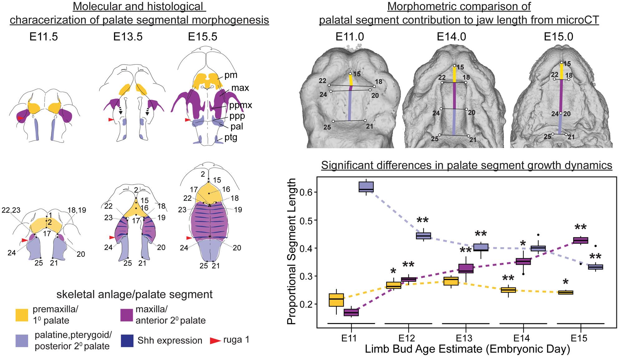
Anterior–posterior (A–P) elongation of the palate is a critical aspect of integrated midfacial morphogenesis. Reciprocal epithelial-mesenchymal interactions drive secondary palate elongation that is coupled to the periodic formation of signaling centers within the rugae growth zone (RGZ). However, the relationship between RGZ-driven morphogenetic processes, the differentiative dynamics of underlying palatal bone mesenchymal precursors, and the segmental organization of the upper jaw has remained enigmatic. A detailed ontogenetic study of these relationships is important because palatal segment growth is a critical aspect of normal midfacial growth, can produce dysmorphology when altered, and is a likely basis for evolutionary differences in upper jaw morphology. We completed a combined whole mount gene expression and morphometric analysis of normal murine palatal segment growth dynamics and resulting upper jaw morphology. Our results demonstrated that the first formed palatal ruga (ruga 1), found just posterior to the RGZ, maintained an association with important nasal, neurovascular and palatal structures throughout early midfacial development. This suggested that these features are positioned at a proximal source of embryonic midfacial directional growth. Our detailed characterization of midfacial morphogenesis revealed a one-to-one relationship between palatal segments and upper jaw bones during the earliest stages of palatal elongation. Growth of the maxillary anlage within the anterior secondary palate is uniquely coupled to RGZ-driven morphogenesis. This may help drive the unequaled proportional elongation of the anterior secondary palate segment prior to palatal shelf fusion. Our results also demonstrated that the future maxillary-palatine suture, approximated by the position of ruga 1 and consistently associated with the palatine anlage, formed predominantly via the posterior differentiation of the maxilla within the expanding anterior secondary palate. Our ontogenetic analysis provides a novel and detailed picture of the earliest spatiotemporal dynamics of intramembranous midfacial skeletal specification and differentiation within the context of the surrounding palatal segment A–P elongation and associated rugae formation.
期刊介绍:
Journal of Anatomy is an international peer-reviewed journal sponsored by the Anatomical Society. The journal publishes original papers, invited review articles and book reviews. Its main focus is to understand anatomy through an analysis of structure, function, development and evolution. Priority will be given to studies of that clearly articulate their relevance to the anatomical community. Focal areas include: experimental studies, contributions based on molecular and cell biology and on the application of modern imaging techniques and papers with novel methods or synthetic perspective on an anatomical system.
Studies that are essentially descriptive anatomy are appropriate only if they communicate clearly a broader functional or evolutionary significance. You must clearly state the broader implications of your work in the abstract.
We particularly welcome submissions in the following areas:
Cell biology and tissue architecture
Comparative functional morphology
Developmental biology
Evolutionary developmental biology
Evolutionary morphology
Functional human anatomy
Integrative vertebrate paleontology
Methodological innovations in anatomical research
Musculoskeletal system
Neuroanatomy and neurodegeneration
Significant advances in anatomical education.


 分享
分享
 求助内容:
求助内容: 应助结果提醒方式:
 扫码关注我们
