Karadi H Sunil Kumar, Floris Van Damme, Ide Van den Borr, Vikas Khanduja, Emmanuel Audenaert, Ajay Malviya
{"title":"Understanding recurrent groin pain following periacetabular osteotomy: assessment of psoas tendon mechanics using discrete element analysis.","authors":"Karadi H Sunil Kumar, Floris Van Damme, Ide Van den Borr, Vikas Khanduja, Emmanuel Audenaert, Ajay Malviya","doi":"10.1093/jhps/hnae020","DOIUrl":null,"url":null,"abstract":"<p><p>Recurrent groin pain following periacetabular osteotomy (PAO) is a challenging problem. The purpose of our study was to evaluate the position and dynamics of the psoas tendon as a potential cause for recurrent groin pain following PAO. A total of 386 PAO procedures, performed between January 2013 and January 2020, were identified from a single surgeon series. Thirteen patients (18 hips) had a psoas tendinopathy, as confirmed with relief of symptoms following a diagnostic injection into the psoas tendon. All patients underwent computed tomography (CT) scans pre- and post-operatively. The data from CT scan was used to manually segment bony structures and create 3D models using Mimics software (Materialise NV). A validated discrete element analysis model using rigid body springs was used to predict psoas tendon movement during hip circumduction and walking. The distance of the iliopsoas tendon to any bony abnormality was calculated. All computational analyses were performed using MATLAB software. Thirteen hips (13/18) showed bony malformations (spurs, hypertrophic callus or delayed union and malunion) secondary to callus at the superior pubic ramus. The mean minimal distance of the iliopsoas tendon to osteotomy site was found to be 13.73 mm (<i>σ</i> = 3.09) for spurs, 10.99 mm (<i>σ</i> = 2.85) for hypertrophic callus and 11.91 mm (<i>σ</i> = 2.55) for canyon type. In normal bony healing, the mean minimal distance was 18.55 mm (<i>σ</i> = 4.11). Using a validated computational modelling technique, this study has demonstrated three different types of malformation around the superior pubic osteotomy site, which are associated with psoas impingement. In all of the cases, the minimal distance of the iliopsoas tendon to the osteotomy site was reduced by 59-74%, as compared with the normal anatomy.</p>","PeriodicalId":48583,"journal":{"name":"Journal of Hip Preservation Surgery","volume":"11 4","pages":"243-250"},"PeriodicalIF":1.1000,"publicationDate":"2024-06-25","publicationTypes":"Journal Article","fieldsOfStudy":null,"isOpenAccess":false,"openAccessPdf":"https://www.ncbi.nlm.nih.gov/pmc/articles/PMC11744472/pdf/","citationCount":"0","resultStr":null,"platform":"Semanticscholar","paperid":null,"PeriodicalName":"Journal of Hip Preservation Surgery","FirstCategoryId":"3","ListUrlMain":"https://doi.org/10.1093/jhps/hnae020","RegionNum":4,"RegionCategory":"医学","ArticlePicture":[],"TitleCN":null,"AbstractTextCN":null,"PMCID":null,"EPubDate":"2024/12/1 0:00:00","PubModel":"eCollection","JCR":"Q3","JCRName":"ORTHOPEDICS","Score":null,"Total":0}
引用次数: 0
Abstract
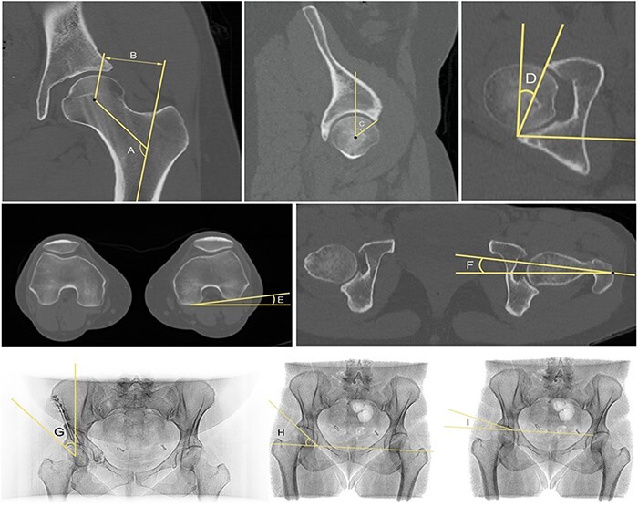
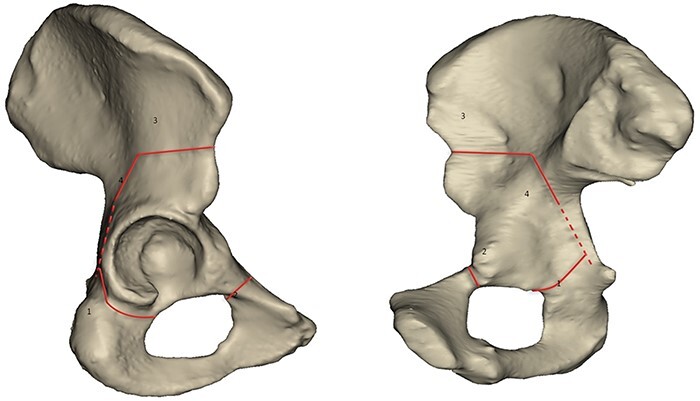
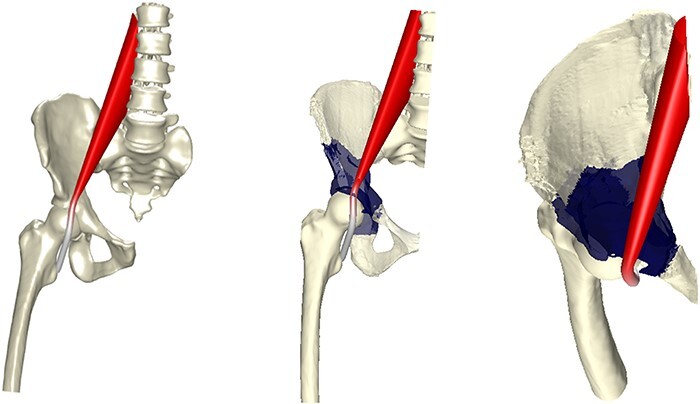
Recurrent groin pain following periacetabular osteotomy (PAO) is a challenging problem. The purpose of our study was to evaluate the position and dynamics of the psoas tendon as a potential cause for recurrent groin pain following PAO. A total of 386 PAO procedures, performed between January 2013 and January 2020, were identified from a single surgeon series. Thirteen patients (18 hips) had a psoas tendinopathy, as confirmed with relief of symptoms following a diagnostic injection into the psoas tendon. All patients underwent computed tomography (CT) scans pre- and post-operatively. The data from CT scan was used to manually segment bony structures and create 3D models using Mimics software (Materialise NV). A validated discrete element analysis model using rigid body springs was used to predict psoas tendon movement during hip circumduction and walking. The distance of the iliopsoas tendon to any bony abnormality was calculated. All computational analyses were performed using MATLAB software. Thirteen hips (13/18) showed bony malformations (spurs, hypertrophic callus or delayed union and malunion) secondary to callus at the superior pubic ramus. The mean minimal distance of the iliopsoas tendon to osteotomy site was found to be 13.73 mm (σ = 3.09) for spurs, 10.99 mm (σ = 2.85) for hypertrophic callus and 11.91 mm (σ = 2.55) for canyon type. In normal bony healing, the mean minimal distance was 18.55 mm (σ = 4.11). Using a validated computational modelling technique, this study has demonstrated three different types of malformation around the superior pubic osteotomy site, which are associated with psoas impingement. In all of the cases, the minimal distance of the iliopsoas tendon to the osteotomy site was reduced by 59-74%, as compared with the normal anatomy.
髋臼周围截骨术后复发性腹股沟疼痛是一个具有挑战性的问题。我们研究的目的是评估腰肌肌腱的位置和动力学作为PAO术后复发性腹股沟疼痛的潜在原因。在2013年1月至2020年1月期间,从单个外科医生系列中确定了386例PAO手术。13例患者(18髋)有腰肌肌腱病变,经诊断性腰大肌肌腱注射后症状缓解。所有患者术前和术后均行CT扫描。CT扫描数据用于手动分割骨骼结构,并使用Mimics软件(Materialise NV)创建3D模型。一个经过验证的离散元分析模型使用刚体弹簧来预测髋关节绕行和行走过程中腰肌肌腱的运动。计算髂腰肌肌腱到任何骨异常的距离。所有计算分析均使用MATLAB软件进行。13髋(13/18)表现为耻骨上支骨痂继发骨畸形(骨刺、肥厚性骨痂或延迟愈合和畸形愈合)。骨刺型髂腰肌肌腱至截骨部位的平均最小距离为13.73 mm (σ = 3.09),肥厚型愈伤组织为10.99 mm (σ = 2.85),峡谷型为11.91 mm (σ = 2.55)。正常骨愈合时,平均最小距离为18.55 mm (σ = 4.11)。使用一种有效的计算建模技术,本研究证明了耻骨上截骨部位周围三种不同类型的畸形,这些畸形与腰肌撞击有关。在所有病例中,与正常解剖相比,髂腰肌肌腱到截骨部位的最小距离减少了59-74%。


 分享
分享
 求助内容:
求助内容: 应助结果提醒方式:
 扫码关注我们
