Michelle Bianchi-de Moraes, Natália Caroline Queiroz Costa, Gabriella Yasmim Santos da Silva, Fernanda Calvo Costa, Fernando Vagner Raldi, Sérgio Lúcio Pereira de Castro Lopes
{"title":"Unveiling degenerative bone changes in the condyle: a texture analysis approach using cone-beam computed tomography.","authors":"Michelle Bianchi-de Moraes, Natália Caroline Queiroz Costa, Gabriella Yasmim Santos da Silva, Fernanda Calvo Costa, Fernando Vagner Raldi, Sérgio Lúcio Pereira de Castro Lopes","doi":"10.1590/acb401325","DOIUrl":null,"url":null,"abstract":"<p><strong>Purpose: </strong>The degenerative joint disease is a temporomandibular disorder. By analysing texture parameters, it becomes possible to characterize and differentiate various tissues, based on their textural properties according to cone-beam computed tomography (CBCT). This study evaluated degenerative diseases in the temporomandibular joint through texture analysis.</p><p><strong>Methods: </strong>Eighty images of the jaw condyle with three types of degenerative diseases, flattening, osteophytes, erosion and control group were analysed, obtained through CBCT. The analyses were carried out through texture analysis with three regions of interest (ROIs) corresponding to specific bone sites. The scans were exported to MaZda software, in which the ROIs were delimited following previously marked contours, and the co-occurrence matrix values were calculated for selected texture analysis parameters.</p><p><strong>Results: </strong>The erosion group showed a significantly different behaviour from the other groups for all analysed parameters.</p><p><strong>Conclusion: </strong>This study highlights the potential of texture analysis in characterizing medullary bone changes in condyles affected by erosion. Texture analysis allows for a more comprehensive assessment of bone condition on CBCT images. These results have implications for early detection and monitoring of degenerative changes in the temporomandibular joint, thus allowing preventive intervention and personalized treatment planning, improving the prognosis of the disease.</p>","PeriodicalId":93850,"journal":{"name":"Acta cirurgica brasileira","volume":"40 ","pages":"e401325"},"PeriodicalIF":1.3000,"publicationDate":"2025-01-13","publicationTypes":"Journal Article","fieldsOfStudy":null,"isOpenAccess":false,"openAccessPdf":"https://www.ncbi.nlm.nih.gov/pmc/articles/PMC11729191/pdf/","citationCount":"0","resultStr":null,"platform":"Semanticscholar","paperid":null,"PeriodicalName":"Acta cirurgica brasileira","FirstCategoryId":"1085","ListUrlMain":"https://doi.org/10.1590/acb401325","RegionNum":0,"RegionCategory":null,"ArticlePicture":[],"TitleCN":null,"AbstractTextCN":null,"PMCID":null,"EPubDate":"2025/1/1 0:00:00","PubModel":"eCollection","JCR":"","JCRName":"","Score":null,"Total":0}
引用次数: 0
Abstract
Purpose: The degenerative joint disease is a temporomandibular disorder. By analysing texture parameters, it becomes possible to characterize and differentiate various tissues, based on their textural properties according to cone-beam computed tomography (CBCT). This study evaluated degenerative diseases in the temporomandibular joint through texture analysis.
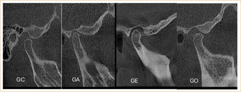
Methods: Eighty images of the jaw condyle with three types of degenerative diseases, flattening, osteophytes, erosion and control group were analysed, obtained through CBCT. The analyses were carried out through texture analysis with three regions of interest (ROIs) corresponding to specific bone sites. The scans were exported to MaZda software, in which the ROIs were delimited following previously marked contours, and the co-occurrence matrix values were calculated for selected texture analysis parameters.
Results: The erosion group showed a significantly different behaviour from the other groups for all analysed parameters.
Conclusion: This study highlights the potential of texture analysis in characterizing medullary bone changes in condyles affected by erosion. Texture analysis allows for a more comprehensive assessment of bone condition on CBCT images. These results have implications for early detection and monitoring of degenerative changes in the temporomandibular joint, thus allowing preventive intervention and personalized treatment planning, improving the prognosis of the disease.


 分享
分享

 求助内容:
求助内容: 应助结果提醒方式:
 扫码关注我们
