Jae Hyun Kim, Myung Ho Cho, Ji Hoon Ban, Sun Hee Lee, Jong Soo Lee
{"title":"Ocular Manifestations of Immune Reconstitution Inflammatory Syndrome in HIV after Highly Active Antiretroviral Therapy: Clinical Use of CD8+ T cell.","authors":"Jae Hyun Kim, Myung Ho Cho, Ji Hoon Ban, Sun Hee Lee, Jong Soo Lee","doi":"10.3341/kjo.2024.0133","DOIUrl":null,"url":null,"abstract":"<p><strong>Purpose: </strong>To investigate ocular manifestation of immune reconstitution inflammatory syndrome (IRIS) in HIV patients after starting highly active antiretroviral therapy (HAART) and its relationship to T cell immunity.</p><p><strong>Methods: </strong>HIV patients with ocular IRIS after HAART were retrospectively reviewed. Clinical presentations with previous opportunistic infection, duration from initiation of HAART to IRIS, blood CD4+, CD8+ T cell count, and HIV RNA copies before HAART and at IRIS were analyzed.</p><p><strong>Results: </strong>Among 19 patients (27 eyes) included, the most common previous opportunistic infection was cytomegalovirus (17 patients, 89.5%) followed by tuberculosis choroiditis (2 patients, 10.5%). The clinical manifestations included vitritis (20 eyes, 74.0%), retinitis (14 eyes, 51.9%), and anterior uveitis (5 eyes, 18.5%). The median duration from initiation of HAART to IRIS was 70 days. CD4+ T cell count before HAART increased at IRIS (p < 0.001). CD8+ T cell count before HAART was negatively correlated with duration from HAART to IRIS (p < 0.001). The cutoff value of CD8+ T cell count for discerning early or late onset of ocular IRIS was 258/mm3 (p = 0.001). When divided into two groups by CD8+ T cell count of 258/mm3, 90% patients with CD8+ T cell count higher than 258/mm3 before HAART developed ocular IRIS within 70 days.</p><p><strong>Conclusions: </strong>There was a negative relationship between CD8+ T cell count before HAART and duration from HAART to ocular IRIS. Ocular IRIS with higher CD8+ T cell count before HAART developed earlier after HAART initiation compared to those with lower CD8+ T cell count.</p>","PeriodicalId":101356,"journal":{"name":"Korean journal of ophthalmology : KJO","volume":" ","pages":"71-79"},"PeriodicalIF":0.0000,"publicationDate":"2025-02-01","publicationTypes":"Journal Article","fieldsOfStudy":null,"isOpenAccess":false,"openAccessPdf":"https://www.ncbi.nlm.nih.gov/pmc/articles/PMC11856080/pdf/","citationCount":"0","resultStr":null,"platform":"Semanticscholar","paperid":null,"PeriodicalName":"Korean journal of ophthalmology : KJO","FirstCategoryId":"1085","ListUrlMain":"https://doi.org/10.3341/kjo.2024.0133","RegionNum":0,"RegionCategory":null,"ArticlePicture":[],"TitleCN":null,"AbstractTextCN":null,"PMCID":null,"EPubDate":"2025/1/20 0:00:00","PubModel":"Epub","JCR":"","JCRName":"","Score":null,"Total":0}
引用次数: 0
Abstract
Purpose: To investigate ocular manifestation of immune reconstitution inflammatory syndrome (IRIS) in HIV patients after starting highly active antiretroviral therapy (HAART) and its relationship to T cell immunity.
Methods: HIV patients with ocular IRIS after HAART were retrospectively reviewed. Clinical presentations with previous opportunistic infection, duration from initiation of HAART to IRIS, blood CD4+, CD8+ T cell count, and HIV RNA copies before HAART and at IRIS were analyzed.
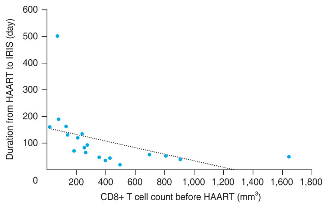
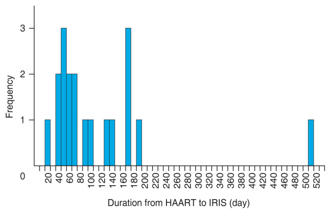
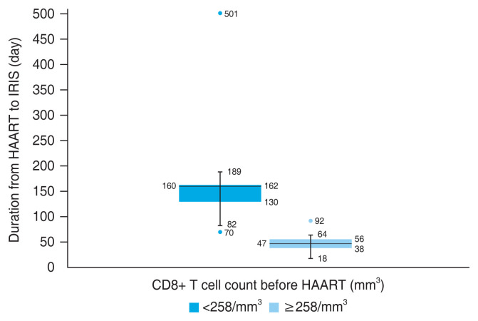
Results: Among 19 patients (27 eyes) included, the most common previous opportunistic infection was cytomegalovirus (17 patients, 89.5%) followed by tuberculosis choroiditis (2 patients, 10.5%). The clinical manifestations included vitritis (20 eyes, 74.0%), retinitis (14 eyes, 51.9%), and anterior uveitis (5 eyes, 18.5%). The median duration from initiation of HAART to IRIS was 70 days. CD4+ T cell count before HAART increased at IRIS (p < 0.001). CD8+ T cell count before HAART was negatively correlated with duration from HAART to IRIS (p < 0.001). The cutoff value of CD8+ T cell count for discerning early or late onset of ocular IRIS was 258/mm3 (p = 0.001). When divided into two groups by CD8+ T cell count of 258/mm3, 90% patients with CD8+ T cell count higher than 258/mm3 before HAART developed ocular IRIS within 70 days.
Conclusions: There was a negative relationship between CD8+ T cell count before HAART and duration from HAART to ocular IRIS. Ocular IRIS with higher CD8+ T cell count before HAART developed earlier after HAART initiation compared to those with lower CD8+ T cell count.


 分享
分享

 求助内容:
求助内容: 应助结果提醒方式:
 扫码关注我们
