Marcin Inglot, Patryk Pozowski, Paula Misiak, Katarzyna Fleischer-Stępniewska, Łukasz Lewandowski, Mateusz Bilski, Urszula Zaleska-Dorobisz
{"title":"Evaluation of liver fibrosis in HCV-infected patients using two-dimensional shear-wave elastography (2D-SWE) before and after antiviral treatment.","authors":"Marcin Inglot, Patryk Pozowski, Paula Misiak, Katarzyna Fleischer-Stępniewska, Łukasz Lewandowski, Mateusz Bilski, Urszula Zaleska-Dorobisz","doi":"10.15557/jou.2024.0038","DOIUrl":null,"url":null,"abstract":"<p><strong>Aim: </strong>Chronic hepatitis C virus infections can lead to liver fibrosis. Appropriate treatment of chronic hepatitis C may result in significant fibrosis reversal. The best method to assess liver fibrosis is an invasive hepatic biopsy. Among non-invasive options, one of the most recent methods is two-dimensional shearwave elastography, which allows real-time visualization of liver stiffness. The purpose of this study was to analyze changes in liver fibrosis among patients with hepatitis C virus receiving direct-acting antiviral therapy.</p><p><strong>Material and methods: </strong>Five different elastographic measurements in kilopascals were performed in a group of 50 patients before direct-acting antiviral treatment, at the end of treatment, and 24 weeks after the end of treatment, using an Aixplorer® (Supersonic Imagine, France) ultrasound device. The results were correlated with biochemical serum tests, specifically the Fibrosis-4 and AspAT-to-platelet ratio indices.</p><p><strong>Results: </strong>Time-dependent alterations of all of the parameters were observed, including a significant decrease in liver stiffness in comparison to baseline values (before treatment). A moderate correlation between liver stiffness measurement values and both Fibrosis-4 and AspAT-to-platelet ratio indices was observed. Interestingly, only liver stiffness and blood platelet count changed over time, regardless of the sex and age of the patient.</p><p><strong>Conclusions: </strong>Two-dimensional shear-wave elastography combined with non-invasive serologic tests like Fibrosis-4 and AspAT-to-platelet ratio indices is a sufficient tool for evaluating liver fibrosis regression during and after direct-acting antiviral therapy.</p>","PeriodicalId":45612,"journal":{"name":"Journal of Ultrasonography","volume":"24 99","pages":"1-10"},"PeriodicalIF":1.5000,"publicationDate":"2024-12-31","publicationTypes":"Journal Article","fieldsOfStudy":null,"isOpenAccess":false,"openAccessPdf":"https://www.ncbi.nlm.nih.gov/pmc/articles/PMC11755404/pdf/","citationCount":"0","resultStr":null,"platform":"Semanticscholar","paperid":null,"PeriodicalName":"Journal of Ultrasonography","FirstCategoryId":"1085","ListUrlMain":"https://doi.org/10.15557/jou.2024.0038","RegionNum":0,"RegionCategory":null,"ArticlePicture":[],"TitleCN":null,"AbstractTextCN":null,"PMCID":null,"EPubDate":"2024/12/1 0:00:00","PubModel":"eCollection","JCR":"Q3","JCRName":"RADIOLOGY, NUCLEAR MEDICINE & MEDICAL IMAGING","Score":null,"Total":0}
引用次数: 0
Abstract
Aim: Chronic hepatitis C virus infections can lead to liver fibrosis. Appropriate treatment of chronic hepatitis C may result in significant fibrosis reversal. The best method to assess liver fibrosis is an invasive hepatic biopsy. Among non-invasive options, one of the most recent methods is two-dimensional shearwave elastography, which allows real-time visualization of liver stiffness. The purpose of this study was to analyze changes in liver fibrosis among patients with hepatitis C virus receiving direct-acting antiviral therapy.
Material and methods: Five different elastographic measurements in kilopascals were performed in a group of 50 patients before direct-acting antiviral treatment, at the end of treatment, and 24 weeks after the end of treatment, using an Aixplorer® (Supersonic Imagine, France) ultrasound device. The results were correlated with biochemical serum tests, specifically the Fibrosis-4 and AspAT-to-platelet ratio indices.
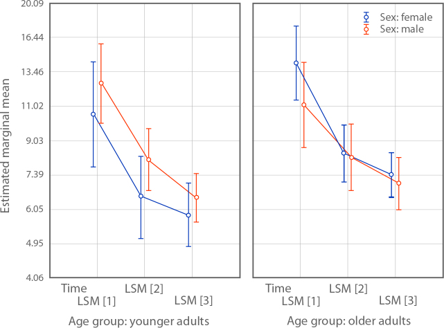
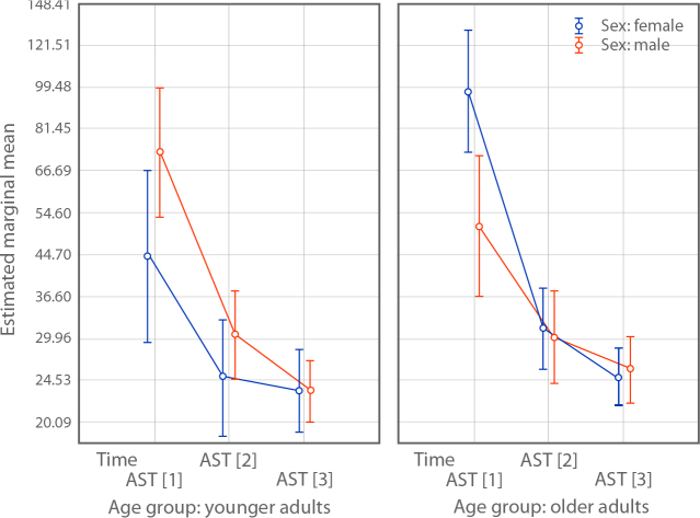
Results: Time-dependent alterations of all of the parameters were observed, including a significant decrease in liver stiffness in comparison to baseline values (before treatment). A moderate correlation between liver stiffness measurement values and both Fibrosis-4 and AspAT-to-platelet ratio indices was observed. Interestingly, only liver stiffness and blood platelet count changed over time, regardless of the sex and age of the patient.
Conclusions: Two-dimensional shear-wave elastography combined with non-invasive serologic tests like Fibrosis-4 and AspAT-to-platelet ratio indices is a sufficient tool for evaluating liver fibrosis regression during and after direct-acting antiviral therapy.


 分享
分享
 求助内容:
求助内容: 应助结果提醒方式:
 扫码关注我们
