Matias Leonardo Cullari, Juan Pablo Taleb, Lucio Gutierrez, Facundo Martín Aguirre, Santiago Alejandro Aguer, Ruy Lloyd, Glenda Ernst
{"title":"Indirect Decompression in Lumbar Degenerative Pathology: Analysis of Imaging Changes at 48 Hours with One-year Follow-up.","authors":"Matias Leonardo Cullari, Juan Pablo Taleb, Lucio Gutierrez, Facundo Martín Aguirre, Santiago Alejandro Aguer, Ruy Lloyd, Glenda Ernst","doi":"10.22038/ABJS.2024.79458.3637","DOIUrl":null,"url":null,"abstract":"<p><strong>Objectives: </strong>Investigate the immediate resonance magnetic image changes undergone by the lumbar canal after indirect decompression and compare them at one-year post-intervention. We also investigate the clinical outcome of indirect decompression at one-year follow-up.</p><p><strong>Methods: </strong>Imaging changes in patients who underwent indirect lumbar decompression and percutaneous posterior fixation were analyzed with one-year follow-up. Radiographic measurements were performed preoperatively and postoperatively (at one year), and the area of lumbar canal occupation and yellow ligament by nuclear magnetic resonance was compared preoperatively, at 48 hours post-surgery, and at one year. Radiographic measurements included disc height, foraminal height, total lumbar lordosis, and segmental lordosis. The VAS lumbar and lower limb scales and the Oswestry Disability Index (ODI) were used to assess clinical outcomes.</p><p><strong>Results: </strong>A total of 21 male and 23 female patients underwent indirect decompression at 64 lumbar levels. A significant improvement was observed in the clinical evaluation of all patients' post-surgery (p < 0.001) in all radiographic parameters. There was an immediate increase in the lumbar canal at 48 hours (p < 0.001), which continued to increase at one year post-intervention (p < 0.05). The yellow ligament occupation area decreased at 48 hours (p < 0.001) and continued to decrease until one year (p < 0.01). Four complications were recorded, one of which was a posterior tract infection requiring open decompression.</p><p><strong>Conclusion: </strong>Indirect decompression for degenerative lumbar disease provided successful clinical outcomes, including indirect expansion of the dural sac at 48 hours post-procedure, with progressive increase in the lumbar canal area at one-year follow-up.</p>","PeriodicalId":46704,"journal":{"name":"Archives of Bone and Joint Surgery-ABJS","volume":"12 11","pages":"779-788"},"PeriodicalIF":1.8000,"publicationDate":"2024-01-01","publicationTypes":"Journal Article","fieldsOfStudy":null,"isOpenAccess":false,"openAccessPdf":"https://www.ncbi.nlm.nih.gov/pmc/articles/PMC11756543/pdf/","citationCount":"0","resultStr":null,"platform":"Semanticscholar","paperid":null,"PeriodicalName":"Archives of Bone and Joint Surgery-ABJS","FirstCategoryId":"1085","ListUrlMain":"https://doi.org/10.22038/ABJS.2024.79458.3637","RegionNum":0,"RegionCategory":null,"ArticlePicture":[],"TitleCN":null,"AbstractTextCN":null,"PMCID":null,"EPubDate":"","PubModel":"","JCR":"Q3","JCRName":"ORTHOPEDICS","Score":null,"Total":0}
引用次数: 0
Abstract
Objectives: Investigate the immediate resonance magnetic image changes undergone by the lumbar canal after indirect decompression and compare them at one-year post-intervention. We also investigate the clinical outcome of indirect decompression at one-year follow-up.
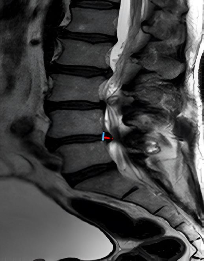
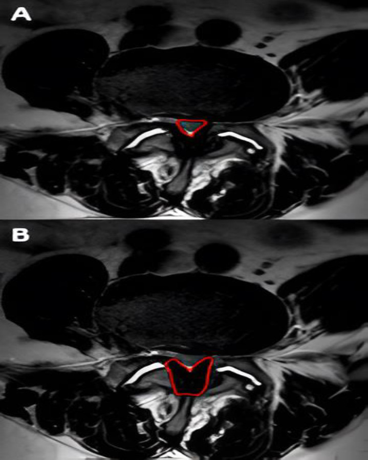
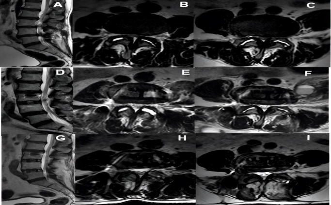
Methods: Imaging changes in patients who underwent indirect lumbar decompression and percutaneous posterior fixation were analyzed with one-year follow-up. Radiographic measurements were performed preoperatively and postoperatively (at one year), and the area of lumbar canal occupation and yellow ligament by nuclear magnetic resonance was compared preoperatively, at 48 hours post-surgery, and at one year. Radiographic measurements included disc height, foraminal height, total lumbar lordosis, and segmental lordosis. The VAS lumbar and lower limb scales and the Oswestry Disability Index (ODI) were used to assess clinical outcomes.
Results: A total of 21 male and 23 female patients underwent indirect decompression at 64 lumbar levels. A significant improvement was observed in the clinical evaluation of all patients' post-surgery (p < 0.001) in all radiographic parameters. There was an immediate increase in the lumbar canal at 48 hours (p < 0.001), which continued to increase at one year post-intervention (p < 0.05). The yellow ligament occupation area decreased at 48 hours (p < 0.001) and continued to decrease until one year (p < 0.01). Four complications were recorded, one of which was a posterior tract infection requiring open decompression.
Conclusion: Indirect decompression for degenerative lumbar disease provided successful clinical outcomes, including indirect expansion of the dural sac at 48 hours post-procedure, with progressive increase in the lumbar canal area at one-year follow-up.
期刊介绍:
The Archives of Bone and Joint Surgery (ABJS) aims to encourage a better understanding of all aspects of Orthopedic Sciences. The journal accepts scientific papers including original research, review article, short communication, case report, and letter to the editor in all fields of bone, joint, musculoskeletal surgery and related researches. The Archives of Bone and Joint Surgery (ABJS) will publish papers in all aspects of today`s modern orthopedic sciences including: Arthroscopy, Arthroplasty, Sport Medicine, Reconstruction, Hand and Upper Extremity, Pediatric Orthopedics, Spine, Trauma, Foot and Ankle, Tumor, Joint Rheumatic Disease, Skeletal Imaging, Orthopedic Physical Therapy, Rehabilitation, Orthopedic Basic Sciences (Biomechanics, Biotechnology, Biomaterial..).


 分享
分享
 求助内容:
求助内容: 应助结果提醒方式:
 扫码关注我们
