Nicholas C. Morano, Yicheng Guo, Jordan E. Becker, Zhiteng Li, Jian Yu, David D. Ho, Lawrence Shapiro, Peter D. Kwong
{"title":"Structure of a zoonotic H5N1 hemagglutinin reveals a receptor-binding site occupied by an auto-glycan","authors":"Nicholas C. Morano, Yicheng Guo, Jordan E. Becker, Zhiteng Li, Jian Yu, David D. Ho, Lawrence Shapiro, Peter D. Kwong","doi":"10.1016/j.str.2025.01.001","DOIUrl":null,"url":null,"abstract":"Highly pathogenic avian influenza has spilled into many mammals, most notably cows and poultry, with several dozen human breakthrough infections. Zoonotic crossovers, with hemagglutinins mutated to enhance viral ability to use human α2-6-linked sialic acid receptors versus avian α2-3-linked ones, highlight the pandemic risk. To gain insight into these crossovers, we determined the cryoelectron microscopy (cryo-EM) structure of the hemagglutinin from the zoonotic H5N1 A/Texas/37/2024 strain (clade 2.3.4.4b) in complex with a previously reported neutralizing antibody. Surprisingly, we found that the receptor-binding site of this H5N1 hemagglutinin was already occupied by an α2-3-linked sialic acid and that this glycan emanated from asparagine N169 of a neighboring protomer on hemagglutinin itself. This structure thus highlights recognition by influenza hemagglutinin of an “auto”-α2-3-linked sialic acid from N169, an <em>N</em>-linked glycan conserved in 95% of H5 strains, and adds “auto-glycan recognition,” which may play a role in viral dispersal, to the complexities surrounding H5N1 zoonosis.","PeriodicalId":22168,"journal":{"name":"Structure","volume":"31 1","pages":""},"PeriodicalIF":4.3000,"publicationDate":"2025-01-29","publicationTypes":"Journal Article","fieldsOfStudy":null,"isOpenAccess":false,"openAccessPdf":"","citationCount":"0","resultStr":null,"platform":"Semanticscholar","paperid":null,"PeriodicalName":"Structure","FirstCategoryId":"99","ListUrlMain":"https://doi.org/10.1016/j.str.2025.01.001","RegionNum":2,"RegionCategory":"生物学","ArticlePicture":[],"TitleCN":null,"AbstractTextCN":null,"PMCID":null,"EPubDate":"","PubModel":"","JCR":"Q2","JCRName":"BIOCHEMISTRY & MOLECULAR BIOLOGY","Score":null,"Total":0}
引用次数: 0
Abstract
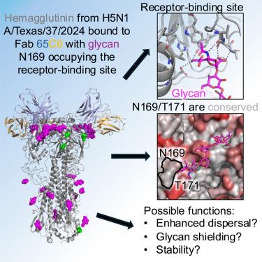
Highly pathogenic avian influenza has spilled into many mammals, most notably cows and poultry, with several dozen human breakthrough infections. Zoonotic crossovers, with hemagglutinins mutated to enhance viral ability to use human α2-6-linked sialic acid receptors versus avian α2-3-linked ones, highlight the pandemic risk. To gain insight into these crossovers, we determined the cryoelectron microscopy (cryo-EM) structure of the hemagglutinin from the zoonotic H5N1 A/Texas/37/2024 strain (clade 2.3.4.4b) in complex with a previously reported neutralizing antibody. Surprisingly, we found that the receptor-binding site of this H5N1 hemagglutinin was already occupied by an α2-3-linked sialic acid and that this glycan emanated from asparagine N169 of a neighboring protomer on hemagglutinin itself. This structure thus highlights recognition by influenza hemagglutinin of an “auto”-α2-3-linked sialic acid from N169, an N-linked glycan conserved in 95% of H5 strains, and adds “auto-glycan recognition,” which may play a role in viral dispersal, to the complexities surrounding H5N1 zoonosis.
期刊介绍:
Structure aims to publish papers of exceptional interest in the field of structural biology. The journal strives to be essential reading for structural biologists, as well as biologists and biochemists that are interested in macromolecular structure and function. Structure strongly encourages the submission of manuscripts that present structural and molecular insights into biological function and mechanism. Other reports that address fundamental questions in structural biology, such as structure-based examinations of protein evolution, folding, and/or design, will also be considered. We will consider the application of any method, experimental or computational, at high or low resolution, to conduct structural investigations, as long as the method is appropriate for the biological, functional, and mechanistic question(s) being addressed. Likewise, reports describing single-molecule analysis of biological mechanisms are welcome.
In general, the editors encourage submission of experimental structural studies that are enriched by an analysis of structure-activity relationships and will not consider studies that solely report structural information unless the structure or analysis is of exceptional and broad interest. Studies reporting only homology models, de novo models, or molecular dynamics simulations are also discouraged unless the models are informed by or validated by novel experimental data; rationalization of a large body of existing experimental evidence and making testable predictions based on a model or simulation is often not considered sufficient.


 分享
分享
 求助内容:
求助内容: 应助结果提醒方式:
 扫码关注我们
