Emily S. Smith-Pillet, Ramya Billur, Marie-France Langelier, Tanaji T. Talele, John M. Pascal, Ben E. Black
{"title":"A PARP2 active site helix melts to permit DNA damage-induced enzymatic activation","authors":"Emily S. Smith-Pillet, Ramya Billur, Marie-France Langelier, Tanaji T. Talele, John M. Pascal, Ben E. Black","doi":"10.1016/j.molcel.2025.01.004","DOIUrl":null,"url":null,"abstract":"Poly(ADP-ribose) polymerase 1 (PARP1) and PARP2 recognize DNA breaks immediately upon their formation, generate a burst of local PARylation to signal their location, and are co-targeted by all current FDA-approved forms of PARP inhibitors (PARPi) used in the cancer clinic. Recent evidence indicates that the same PARPi molecules impact PARP2 differently from PARP1, raising the possibility that allosteric activation may also differ. We find that, unlike for PARP1, destabilization of the autoinhibitory domain of PARP2 is insufficient for DNA damage-induced catalytic activation. Rather, PARP2 activation requires further unfolding of an active site helix. In contrast, the corresponding helix in PARP1 only transiently forms, even prior to engaging DNA. Only one clinical PARPi, Olaparib, stabilizes the PARP2 active site helix, representing a structural feature with the potential to discriminate small molecule inhibitors. Collectively, our findings reveal unanticipated differences in local structure and changes in activation-coupled backbone dynamics between human PARP1 and PARP2.","PeriodicalId":18950,"journal":{"name":"Molecular Cell","volume":"36 1","pages":""},"PeriodicalIF":16.6000,"publicationDate":"2025-01-30","publicationTypes":"Journal Article","fieldsOfStudy":null,"isOpenAccess":false,"openAccessPdf":"","citationCount":"0","resultStr":null,"platform":"Semanticscholar","paperid":null,"PeriodicalName":"Molecular Cell","FirstCategoryId":"99","ListUrlMain":"https://doi.org/10.1016/j.molcel.2025.01.004","RegionNum":1,"RegionCategory":"生物学","ArticlePicture":[],"TitleCN":null,"AbstractTextCN":null,"PMCID":null,"EPubDate":"","PubModel":"","JCR":"Q1","JCRName":"BIOCHEMISTRY & MOLECULAR BIOLOGY","Score":null,"Total":0}
引用次数: 0
Abstract
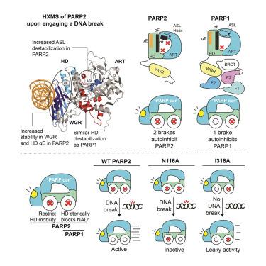
Poly(ADP-ribose) polymerase 1 (PARP1) and PARP2 recognize DNA breaks immediately upon their formation, generate a burst of local PARylation to signal their location, and are co-targeted by all current FDA-approved forms of PARP inhibitors (PARPi) used in the cancer clinic. Recent evidence indicates that the same PARPi molecules impact PARP2 differently from PARP1, raising the possibility that allosteric activation may also differ. We find that, unlike for PARP1, destabilization of the autoinhibitory domain of PARP2 is insufficient for DNA damage-induced catalytic activation. Rather, PARP2 activation requires further unfolding of an active site helix. In contrast, the corresponding helix in PARP1 only transiently forms, even prior to engaging DNA. Only one clinical PARPi, Olaparib, stabilizes the PARP2 active site helix, representing a structural feature with the potential to discriminate small molecule inhibitors. Collectively, our findings reveal unanticipated differences in local structure and changes in activation-coupled backbone dynamics between human PARP1 and PARP2.
期刊介绍:
Molecular Cell is a companion to Cell, the leading journal of biology and the highest-impact journal in the world. Launched in December 1997 and published monthly. Molecular Cell is dedicated to publishing cutting-edge research in molecular biology, focusing on fundamental cellular processes. The journal encompasses a wide range of topics, including DNA replication, recombination, and repair; Chromatin biology and genome organization; Transcription; RNA processing and decay; Non-coding RNA function; Translation; Protein folding, modification, and quality control; Signal transduction pathways; Cell cycle and checkpoints; Cell death; Autophagy; Metabolism.


 分享
分享
 求助内容:
求助内容: 应助结果提醒方式:
 扫码关注我们
