High-resolution 0.25 mm Detector CT Has Limited Impact on Right Adrenal Vein Detectability in Preprocedural Contrast Enhanced CT for Adrenal Venous Sampling.
{"title":"High-resolution 0.25 mm Detector CT Has Limited Impact on Right Adrenal Vein Detectability in Preprocedural Contrast Enhanced CT for Adrenal Venous Sampling.","authors":"Hiroyuki Morisaka, Akira Imaizumi, Tihan Wumu, Takanori Ii, Takuji Araki, Hiroshi Onishi","doi":"10.1097/RCT.0000000000001727","DOIUrl":null,"url":null,"abstract":"<p><strong>Objective: </strong>This study aims to identify factors associated with the detectability of the right adrenal vein (RAV) on preoperative contrast-enhanced CT scans of adrenal venous sampling (AVS) in the era of high-resolution CT (HRCT).</p><p><strong>Materials and methods: </strong>In this retrospective study, 36 patients (15 men and 21 women; mean age, 56 y) who underwent preoperative contrast-enhanced CT [11 patients in HRCT with 0.25 mm detector matrix (Cannon Medical Systems) and 25 patients in conventional multidetector CT with 0.5 mm matrix] were included. A contrast agent dose of 600 mgI/kg was injected, and CT images were acquired at a fixed scan delay of 50 and 80 seconds. Adrenal venography and venous sampling were performed for the diagnosis of suspected primary hyperaldosteronism. The qualitative detectability of RAV on preoperative CT was assessed with adrenal venography as a reference. Clinical and imaging factors associated with a good detectability of RAV were analyzed via regression analysis. Optimal acquisition timing was assessed by analyzing the time-intensity curve and contrast enhancement pattern of the inferior vena cava using CT data from a separate cohort (n=5).</p><p><strong>Results: </strong>The qualitative detectability of RAV was deemed good in 15 patients and poor in 21 patients. Regression analysis revealed that only heterogeneous enhancement of inferior vena cava with bolus high attenuation, corresponding to an optimal acquisition timing from time-intensity curve analysis, was associated with a good detectability of RAV (odds ratio, 5.06). The use of HRCT was not statistically significant.</p><p><strong>Conclusions: </strong>Optimal acquisition timing is a crucial factor for the detectability of RAV in preprocedural CT for AVS, while high-resolution 0.25 detector CT appears to have limited significance.</p>","PeriodicalId":15402,"journal":{"name":"Journal of Computer Assisted Tomography","volume":" ","pages":"745-750"},"PeriodicalIF":1.3000,"publicationDate":"2025-09-01","publicationTypes":"Journal Article","fieldsOfStudy":null,"isOpenAccess":false,"openAccessPdf":"https://www.ncbi.nlm.nih.gov/pmc/articles/PMC12430825/pdf/","citationCount":"0","resultStr":null,"platform":"Semanticscholar","paperid":null,"PeriodicalName":"Journal of Computer Assisted Tomography","FirstCategoryId":"3","ListUrlMain":"https://doi.org/10.1097/RCT.0000000000001727","RegionNum":4,"RegionCategory":"医学","ArticlePicture":[],"TitleCN":null,"AbstractTextCN":null,"PMCID":null,"EPubDate":"2025/1/27 0:00:00","PubModel":"Epub","JCR":"Q4","JCRName":"RADIOLOGY, NUCLEAR MEDICINE & MEDICAL IMAGING","Score":null,"Total":0}
引用次数: 0
Abstract
Objective: This study aims to identify factors associated with the detectability of the right adrenal vein (RAV) on preoperative contrast-enhanced CT scans of adrenal venous sampling (AVS) in the era of high-resolution CT (HRCT).
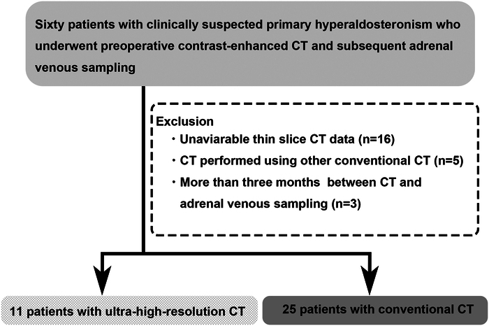
Materials and methods: In this retrospective study, 36 patients (15 men and 21 women; mean age, 56 y) who underwent preoperative contrast-enhanced CT [11 patients in HRCT with 0.25 mm detector matrix (Cannon Medical Systems) and 25 patients in conventional multidetector CT with 0.5 mm matrix] were included. A contrast agent dose of 600 mgI/kg was injected, and CT images were acquired at a fixed scan delay of 50 and 80 seconds. Adrenal venography and venous sampling were performed for the diagnosis of suspected primary hyperaldosteronism. The qualitative detectability of RAV on preoperative CT was assessed with adrenal venography as a reference. Clinical and imaging factors associated with a good detectability of RAV were analyzed via regression analysis. Optimal acquisition timing was assessed by analyzing the time-intensity curve and contrast enhancement pattern of the inferior vena cava using CT data from a separate cohort (n=5).
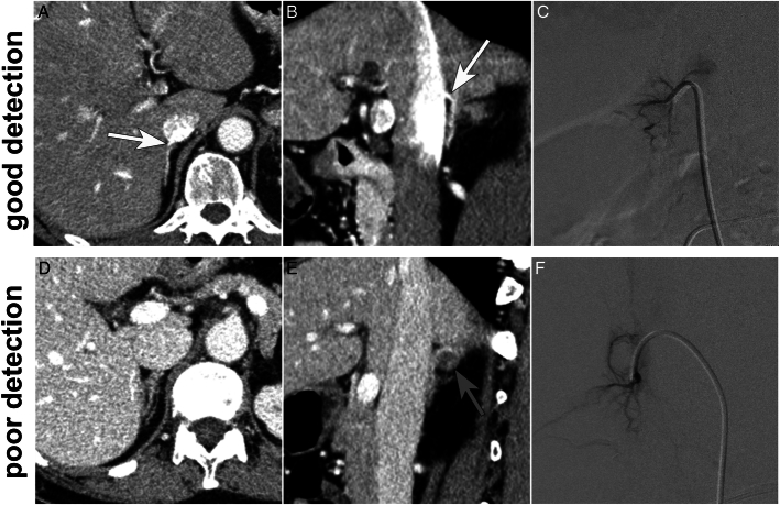
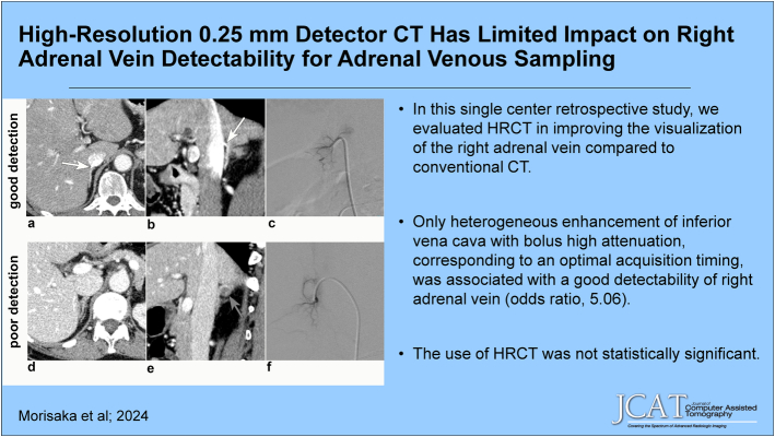
Results: The qualitative detectability of RAV was deemed good in 15 patients and poor in 21 patients. Regression analysis revealed that only heterogeneous enhancement of inferior vena cava with bolus high attenuation, corresponding to an optimal acquisition timing from time-intensity curve analysis, was associated with a good detectability of RAV (odds ratio, 5.06). The use of HRCT was not statistically significant.
Conclusions: Optimal acquisition timing is a crucial factor for the detectability of RAV in preprocedural CT for AVS, while high-resolution 0.25 detector CT appears to have limited significance.
期刊介绍:
The mission of Journal of Computer Assisted Tomography is to showcase the latest clinical and research developments in CT, MR, and closely related diagnostic techniques. We encourage submission of both original research and review articles that have immediate or promissory clinical applications. Topics of special interest include: 1) functional MR and CT of the brain and body; 2) advanced/innovative MRI techniques (diffusion, perfusion, rapid scanning); and 3) advanced/innovative CT techniques (perfusion, multi-energy, dose-reduction, and processing).


 分享
分享
 求助内容:
求助内容: 应助结果提醒方式:
 扫码关注我们
