Ryckie G Wade, Gráinne Bourke, Alexandra M Olaru, Stephen R Williams, David Shelley, Sven Plein, Robert D Bains, James D Bedford, Lucy E Homer Newton, Chye Yew Ng, Laura Parkes, Caroline Lea-Carnall
{"title":"Cortical Neurotransmitters Measured by Magnetic Resonance Spectroscopy Change Following Traumatic Brachial Plexus Injury.","authors":"Ryckie G Wade, Gráinne Bourke, Alexandra M Olaru, Stephen R Williams, David Shelley, Sven Plein, Robert D Bains, James D Bedford, Lucy E Homer Newton, Chye Yew Ng, Laura Parkes, Caroline Lea-Carnall","doi":"10.1055/a-2505-5657","DOIUrl":null,"url":null,"abstract":"<p><p><b>Introduction</b> GABA (γ-aminobutyric acid) is the major inhibitory neurotransmitter in the brain. In response to injury within the central nervous system, GABA promotes cortical plasticity and represents a potential pharmacological target to improve functional recovery. However, it is unclear how GABA changes in the brain after traumatic brachial plexus injuries (tBPIs) which represents the rationale for this pilot study. <b>Methods</b> We serially scanned seven males (mean age 42 years [SD 19] without head injury) up to 19 months after tBPIs. T1-weighted images (1-mm isotropic resolution) and J-edited spectra (MEscher-GArwood Point RESolved Spectroscopy [MEGA-PRESS], TE 68 ms, TR 2,000 ms, 2 cm isotropic voxels) were acquired using a MAGNETOM Prisma 3T (Siemens Healthcare, Erlangen, Germany). Data were analyzed in jMRUI blind to clinical information to quantify GABA, creatine plus phosphocreatine (Cr), and N-acetylaspartate (NAA) concentrations. Additionally, gray matter and white matter proportions were assessed using SPECTRIM software. Interhemispheric means were compared using linear methods. Confidence intervals (CIs) were generated to the 95% level. <b>Results</b> Within weeks of injury, the hemisphere representing the injured upper limb had a significantly lower GABA:NAA ratio (mean difference 0.23 [CI 0.06-0.40]) and GABA:Cr ratio (mean difference 0.75 [CI 0.24-1.25]) than the uninjured side. There were no interhemispheric differences in NAA:Cr. By 12 months post-injury, interhemispheric differences in metabolite concentrations equalized. There was no difference in the proportion of gray matter, white matter, or cerebrospinal fluid between the injured and uninjured hemispheres. <b>Conclusion</b> After brachial plexus injuries, there are interhemispheric differences in GABA concentrations within the sensory and motor cortex. This represents a potential pharmacological target that warrants further investigation.</p>","PeriodicalId":15280,"journal":{"name":"Journal of Brachial Plexus and Peripheral Nerve Injury","volume":"20 1","pages":"e16-e25"},"PeriodicalIF":1.0000,"publicationDate":"2025-01-28","publicationTypes":"Journal Article","fieldsOfStudy":null,"isOpenAccess":false,"openAccessPdf":"https://www.ncbi.nlm.nih.gov/pmc/articles/PMC11774636/pdf/","citationCount":"0","resultStr":null,"platform":"Semanticscholar","paperid":null,"PeriodicalName":"Journal of Brachial Plexus and Peripheral Nerve Injury","FirstCategoryId":"1085","ListUrlMain":"https://doi.org/10.1055/a-2505-5657","RegionNum":0,"RegionCategory":null,"ArticlePicture":[],"TitleCN":null,"AbstractTextCN":null,"PMCID":null,"EPubDate":"2025/1/1 0:00:00","PubModel":"eCollection","JCR":"Q4","JCRName":"CLINICAL NEUROLOGY","Score":null,"Total":0}
引用次数: 0
Abstract
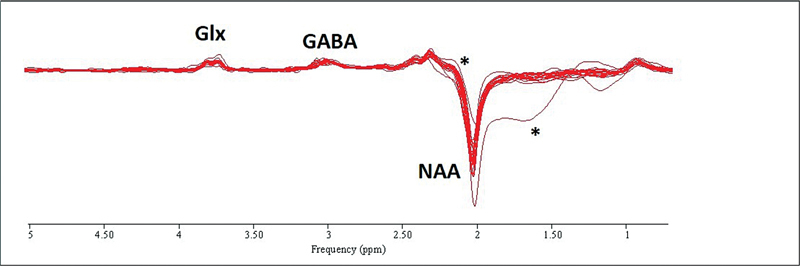
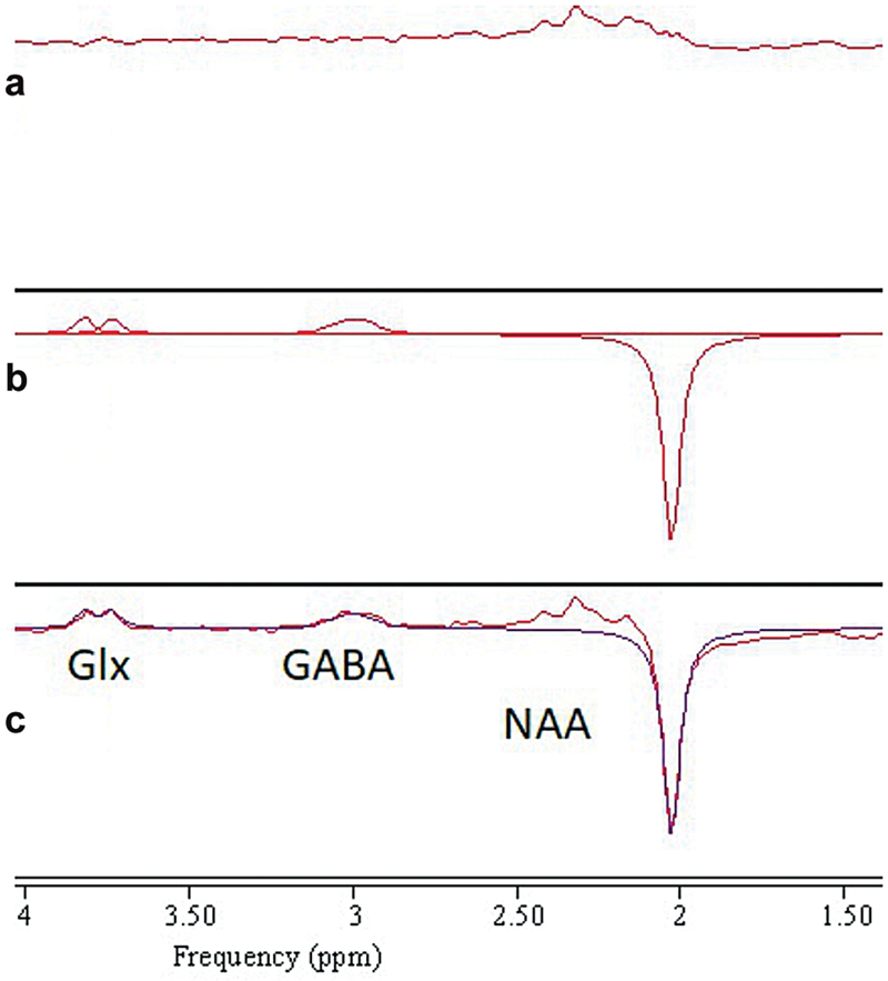
Introduction GABA (γ-aminobutyric acid) is the major inhibitory neurotransmitter in the brain. In response to injury within the central nervous system, GABA promotes cortical plasticity and represents a potential pharmacological target to improve functional recovery. However, it is unclear how GABA changes in the brain after traumatic brachial plexus injuries (tBPIs) which represents the rationale for this pilot study. Methods We serially scanned seven males (mean age 42 years [SD 19] without head injury) up to 19 months after tBPIs. T1-weighted images (1-mm isotropic resolution) and J-edited spectra (MEscher-GArwood Point RESolved Spectroscopy [MEGA-PRESS], TE 68 ms, TR 2,000 ms, 2 cm isotropic voxels) were acquired using a MAGNETOM Prisma 3T (Siemens Healthcare, Erlangen, Germany). Data were analyzed in jMRUI blind to clinical information to quantify GABA, creatine plus phosphocreatine (Cr), and N-acetylaspartate (NAA) concentrations. Additionally, gray matter and white matter proportions were assessed using SPECTRIM software. Interhemispheric means were compared using linear methods. Confidence intervals (CIs) were generated to the 95% level. Results Within weeks of injury, the hemisphere representing the injured upper limb had a significantly lower GABA:NAA ratio (mean difference 0.23 [CI 0.06-0.40]) and GABA:Cr ratio (mean difference 0.75 [CI 0.24-1.25]) than the uninjured side. There were no interhemispheric differences in NAA:Cr. By 12 months post-injury, interhemispheric differences in metabolite concentrations equalized. There was no difference in the proportion of gray matter, white matter, or cerebrospinal fluid between the injured and uninjured hemispheres. Conclusion After brachial plexus injuries, there are interhemispheric differences in GABA concentrations within the sensory and motor cortex. This represents a potential pharmacological target that warrants further investigation.
期刊介绍:
JBPPNI is an open access, peer-reviewed online journal that will encompass all aspects of basic and clinical research findings, in the area of brachial plexus and peripheral nerve injury. Injury in this context refers to congenital, inflammatory, traumatic, degenerative and neoplastic processes, including neurofibromatosis. Papers on diagnostic and imaging aspects of the peripheral nervous system are welcomed as well. The peripheral nervous system is unique in its complexity and scope of influence. There are areas of interest in the anatomy, physiology, metabolism, phylogeny, and limb growth tropism of peripheral nerves.


 分享
分享
 求助内容:
求助内容: 应助结果提醒方式:
 扫码关注我们
