{"title":"Radiological Findings for Distinguishing Between Xanthogranulomatous Cholecystitis and Gallbladder Cancer.","authors":"Ahmet Bozer, Nagihan Durgun","doi":"10.34172/aim.31710","DOIUrl":null,"url":null,"abstract":"<p><strong>Background: </strong>Xanthogranulomatous cholecystitis (XGC) is a rare, chronic gallbladder inflammation often mistaken for gallbladder cancer (GBC) on imaging. Accurate differentiation is vital for appropriate treatment. This study aims to enhance computed tomography (CT) scan diagnostic accuracy for distinguishing XGC from GBC.</p><p><strong>Methods: </strong>This retrospective study included patients diagnosed with XGC and GBC between 2014 and 2023. CT images of 70 patients (16 GBC, 54 XGC) were reviewed. Radiologists assessed CT parameters: gallbladder wall thickening, intramural hypoattenuating nodules, enhancement characteristics, mucosal line continuity, pericholecystic fat stranding, presence of stones, bile duct dilatation, hepatic invasion, invasion to adjacent structures, and lymph node size.</p><p><strong>Results: </strong>Among 70 patients, there were 38 males (54%) and 32 females (46%), with a median age of 62 years. GBC patients were significantly older (median age 72 years) compared to XGC patients (60 years) (<i>P</i>=0.001). Diffuse gallbladder wall thickening was more frequent in XGC (70%) than GBC (12.5%) (<i>P</i><0.001). Continuous mucosal lines and intramural hypoattenuating nodules were more common in XGC (<i>P</i><0.001 and <i>P</i>=0.010, respectively). Intrahepatic bile duct dilatation and invasion to adjacent structures were significantly linked with GBC (<i>P</i><0.001 and <i>P</i>=0.043). Lymph nodes with a short axis>8 mm indicated GBC (<i>P</i><0.001), with a cutoff providing 71.4% sensitivity and 84% specificity (AUC: 0.843, <i>P</i><0.001). CT showed 75% sensitivity (95% CI: 48-93%), 74% specificity (95% CI: 60%-85%), and 74% accuracy (95% CI: 62%-84%).</p><p><strong>Conclusion: </strong>CT imaging can effectively differentiate XGC from GBC, and larger studies can further improve diagnostic accuracy.</p>","PeriodicalId":55469,"journal":{"name":"Archives of Iranian Medicine","volume":"27 12","pages":"674-682"},"PeriodicalIF":1.0000,"publicationDate":"2024-12-01","publicationTypes":"Journal Article","fieldsOfStudy":null,"isOpenAccess":false,"openAccessPdf":"https://www.ncbi.nlm.nih.gov/pmc/articles/PMC11786208/pdf/","citationCount":"0","resultStr":null,"platform":"Semanticscholar","paperid":null,"PeriodicalName":"Archives of Iranian Medicine","FirstCategoryId":"3","ListUrlMain":"https://doi.org/10.34172/aim.31710","RegionNum":4,"RegionCategory":"医学","ArticlePicture":[],"TitleCN":null,"AbstractTextCN":null,"PMCID":null,"EPubDate":"","PubModel":"","JCR":"Q3","JCRName":"MEDICINE, GENERAL & INTERNAL","Score":null,"Total":0}
引用次数: 0
Abstract
Background: Xanthogranulomatous cholecystitis (XGC) is a rare, chronic gallbladder inflammation often mistaken for gallbladder cancer (GBC) on imaging. Accurate differentiation is vital for appropriate treatment. This study aims to enhance computed tomography (CT) scan diagnostic accuracy for distinguishing XGC from GBC.
Methods: This retrospective study included patients diagnosed with XGC and GBC between 2014 and 2023. CT images of 70 patients (16 GBC, 54 XGC) were reviewed. Radiologists assessed CT parameters: gallbladder wall thickening, intramural hypoattenuating nodules, enhancement characteristics, mucosal line continuity, pericholecystic fat stranding, presence of stones, bile duct dilatation, hepatic invasion, invasion to adjacent structures, and lymph node size.
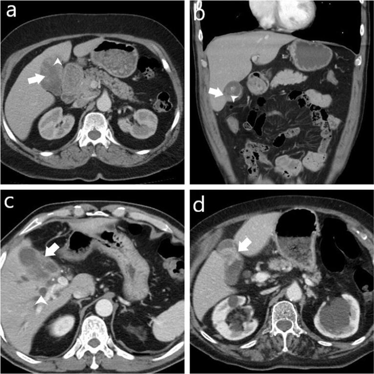
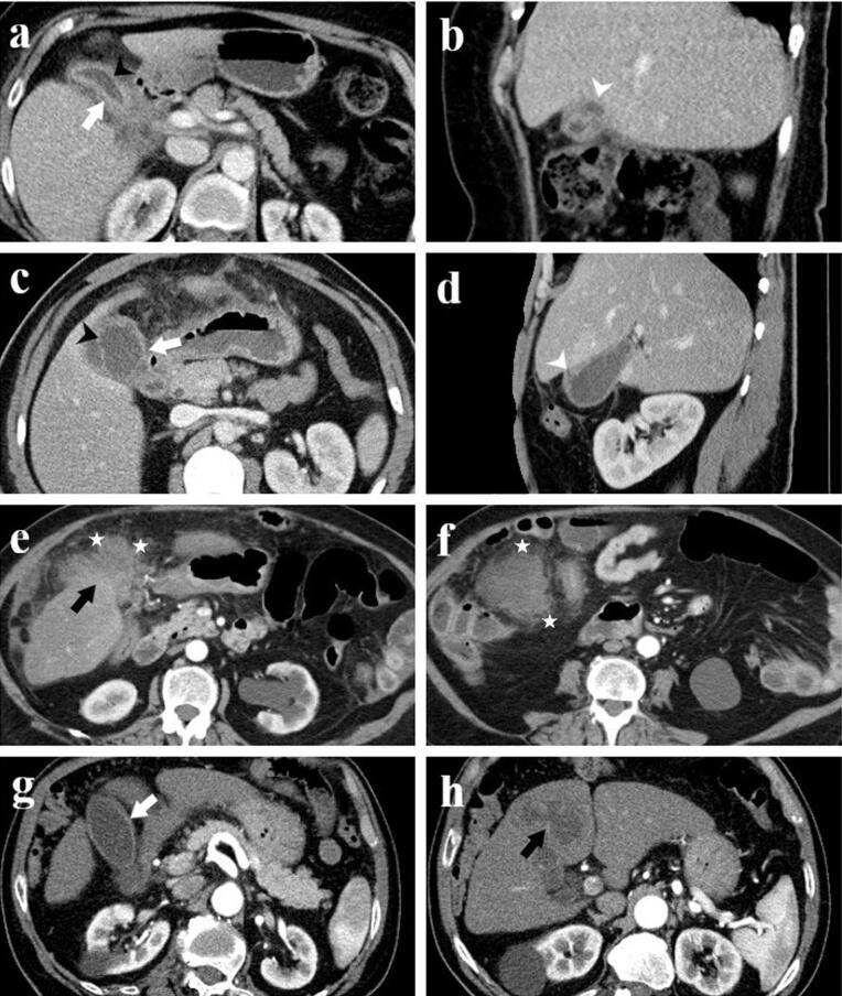
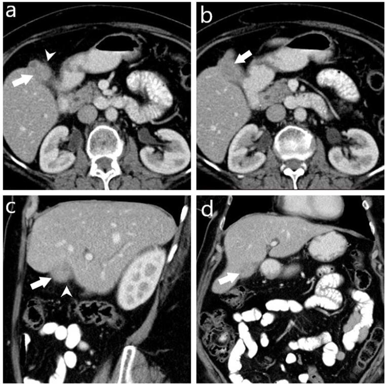
Results: Among 70 patients, there were 38 males (54%) and 32 females (46%), with a median age of 62 years. GBC patients were significantly older (median age 72 years) compared to XGC patients (60 years) (P=0.001). Diffuse gallbladder wall thickening was more frequent in XGC (70%) than GBC (12.5%) (P<0.001). Continuous mucosal lines and intramural hypoattenuating nodules were more common in XGC (P<0.001 and P=0.010, respectively). Intrahepatic bile duct dilatation and invasion to adjacent structures were significantly linked with GBC (P<0.001 and P=0.043). Lymph nodes with a short axis>8 mm indicated GBC (P<0.001), with a cutoff providing 71.4% sensitivity and 84% specificity (AUC: 0.843, P<0.001). CT showed 75% sensitivity (95% CI: 48-93%), 74% specificity (95% CI: 60%-85%), and 74% accuracy (95% CI: 62%-84%).
Conclusion: CT imaging can effectively differentiate XGC from GBC, and larger studies can further improve diagnostic accuracy.
期刊介绍:
Aim and Scope: The Archives of Iranian Medicine (AIM) is a monthly peer-reviewed multidisciplinary medical publication. The journal welcomes contributions particularly relevant to the Middle-East region and publishes biomedical experiences and clinical investigations on prevalent diseases in the region as well as analyses of factors that may modulate the incidence, course, and management of diseases and pertinent medical problems. Manuscripts with didactic orientation and subjects exclusively of local interest will not be considered for publication.The 2016 Impact Factor of "Archives of Iranian Medicine" is 1.20.


 分享
分享
 求助内容:
求助内容: 应助结果提醒方式:
 扫码关注我们
