Jeffrey J. Feng MS , Sophie M. Cannon MD , Stephanie K. Cheok MD , Mark S. Shiroishi MD , Kyle M. Hurth MD, PhD , Anna J. Mathew MD , Gabriel Zada MD , John D. Carmichael MD
{"title":"Successful Multimodal Management of an Aggressive Functional Gonadotropic Pituitary Macroadenoma","authors":"Jeffrey J. Feng MS , Sophie M. Cannon MD , Stephanie K. Cheok MD , Mark S. Shiroishi MD , Kyle M. Hurth MD, PhD , Anna J. Mathew MD , Gabriel Zada MD , John D. Carmichael MD","doi":"10.1016/j.aace.2024.09.003","DOIUrl":null,"url":null,"abstract":"<div><h3>Background/Objective</h3><div>Although most gonadotroph cell–derived pituitary adenomas (PAs) give rise to nonfunctional PAs, hormonally active functional gonadotroph adenomas (FGAs) are exceedingly rare. We present a case of a giant and invasive functional gonadotropic pituitary macroadenoma treated with endoscopic transsphenoidal surgery and subsequent postoperative radiotherapy.</div></div><div><h3>Case Report</h3><div>A 54-year-old man presented with gradually worsening vision over 1 year. Magnetic resonance imaging demonstrated a 5.2-cm sellar and suprasellar mass with cavernous sinus invasion, mass effect on the optic chiasm, and extension into the sphenoid sinus, nasal cavity, and clivus. Preoperative workup was remarkable for erythrocytosis without sleep apnea and increased levels of follicle-stimulating hormone (FSH), luteinizing hormone (LH), prolactin, and testosterone. Immunohistochemistry results following endoscopic transsphenoidal resection confirmed dominant staining for steroidogenic factor-1, FSH, and LH. Postoperatively, the patient’s FSH level decreased, whereas the LH level normalized within 1 week. The free testosterone level normalized at 9 months. The patient underwent radiotherapy for a small amount of residual tumor in the right cavernous sinus and has demonstrated no evidence of disease or hormonal progression.</div></div><div><h3>Discussion</h3><div>There is no consensus on FGA-specific management that differs from the management of nonfunctional PAs; surgery is recommended when vision is impacted. The invasive nature of the tumor presented in this case is rare and limited safe gross total resection, requiring adjuvant radiotherapy.</div></div><div><h3>Conclusion</h3><div>FGAs are rare, and those of similar size and extent of invasion as in our case are even more so. In addition to surgical resection, consideration of adjunct therapies including radiation and multidisciplinary physician involvement are vital in achieving clinical improvement and remission while preventing possible progression and recurrence.</div></div>","PeriodicalId":7051,"journal":{"name":"AACE Clinical Case Reports","volume":"11 1","pages":"Pages 14-17"},"PeriodicalIF":1.2000,"publicationDate":"2025-01-01","publicationTypes":"Journal Article","fieldsOfStudy":null,"isOpenAccess":false,"openAccessPdf":"https://www.ncbi.nlm.nih.gov/pmc/articles/PMC11784608/pdf/","citationCount":"0","resultStr":null,"platform":"Semanticscholar","paperid":null,"PeriodicalName":"AACE Clinical Case Reports","FirstCategoryId":"1085","ListUrlMain":"https://www.sciencedirect.com/science/article/pii/S2376060524001019","RegionNum":0,"RegionCategory":null,"ArticlePicture":[],"TitleCN":null,"AbstractTextCN":null,"PMCID":null,"EPubDate":"","PubModel":"","JCR":"Q3","JCRName":"Medicine","Score":null,"Total":0}
引用次数: 0
Abstract
Background/Objective
Although most gonadotroph cell–derived pituitary adenomas (PAs) give rise to nonfunctional PAs, hormonally active functional gonadotroph adenomas (FGAs) are exceedingly rare. We present a case of a giant and invasive functional gonadotropic pituitary macroadenoma treated with endoscopic transsphenoidal surgery and subsequent postoperative radiotherapy.
Case Report
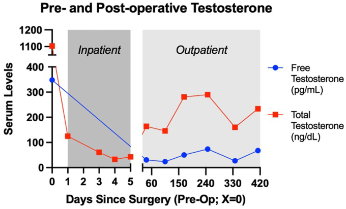
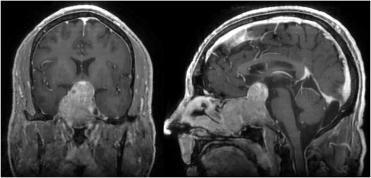
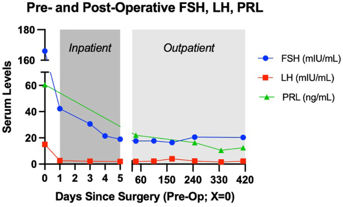
A 54-year-old man presented with gradually worsening vision over 1 year. Magnetic resonance imaging demonstrated a 5.2-cm sellar and suprasellar mass with cavernous sinus invasion, mass effect on the optic chiasm, and extension into the sphenoid sinus, nasal cavity, and clivus. Preoperative workup was remarkable for erythrocytosis without sleep apnea and increased levels of follicle-stimulating hormone (FSH), luteinizing hormone (LH), prolactin, and testosterone. Immunohistochemistry results following endoscopic transsphenoidal resection confirmed dominant staining for steroidogenic factor-1, FSH, and LH. Postoperatively, the patient’s FSH level decreased, whereas the LH level normalized within 1 week. The free testosterone level normalized at 9 months. The patient underwent radiotherapy for a small amount of residual tumor in the right cavernous sinus and has demonstrated no evidence of disease or hormonal progression.
Discussion
There is no consensus on FGA-specific management that differs from the management of nonfunctional PAs; surgery is recommended when vision is impacted. The invasive nature of the tumor presented in this case is rare and limited safe gross total resection, requiring adjuvant radiotherapy.
Conclusion
FGAs are rare, and those of similar size and extent of invasion as in our case are even more so. In addition to surgical resection, consideration of adjunct therapies including radiation and multidisciplinary physician involvement are vital in achieving clinical improvement and remission while preventing possible progression and recurrence.


 分享
分享
 求助内容:
求助内容: 应助结果提醒方式:
 扫码关注我们
