Muhammad Jehanzeb Khan, Zainab Rustam, Faiqa Binte Aamir, Maria Chairez Miranda, Imad Shaikh, Anam Akhlaq, Jiawen Liu, Mandeep Singh, Xiangrong Kong, Peter A Campochiaro
{"title":"Characterization of Macular Fundus Autofluorescence Changes in Patients with Retinitis Pigmentosa.","authors":"Muhammad Jehanzeb Khan, Zainab Rustam, Faiqa Binte Aamir, Maria Chairez Miranda, Imad Shaikh, Anam Akhlaq, Jiawen Liu, Mandeep Singh, Xiangrong Kong, Peter A Campochiaro","doi":"10.1159/000543082","DOIUrl":null,"url":null,"abstract":"<p><strong>Introduction: </strong>The aim of this study was to characterized fundus autofluorescence (AF) changes that occur in the macula of patients with retinitis pigmentosa (RP).</p><p><strong>Methods: </strong>We conducted a case series on 99 patients with RP. Features seen on fundus AF images were evaluated, organized into a grading scheme, and correlated with ellipsoid zone (EZ) width. Patterns of AF changes occurring in the macula and correlation with EZ width.</p><p><strong>Results: </strong>Four primary fundus AF phenotypes were identified: (1) hyperAF arc, (2) hyperAF ring, (3) hyperAF ring with abnormal hyperAF within the ring, and (4) central hyperAF. The second phenotype was most common and had 3 subgroups, hyperAF rings within the macula, those extending outside the macula, and incomplete rings. HyperAF rings were also characterized as narrow or wide with wide rings having a greater amount of hyperAF. Linear Mixed-Effects Model showed mean measured EZ width was significantly greater for phenotype 1 versus each of the other 3 phenotypes (p < 0.01) and for phenotype 2 versus phenotypes 3 and 4 (p < 0.05), and also differed among phenotype 2 subgroups (p < 0.05). Other AF characteristics identified were focal posterior distinct or indistinct hypoAF which sometimes formed complete or incomplete hypoAF rings surrounding a hyperAF ring, diffuse or focal hyperAF outside hyperAF rings, and the amount of encroachment of peripheral hypoAF.</p><p><strong>Conclusions: </strong>A grading scheme for macular AF features in patients with RP identified phenotypes that correlate with stage of disease based upon EZ width. Longitudinal studies are needed to test whether presumed early AF phenotypes evolve into later phenotypes. Use of the grading scheme for patient populations in interventional trials could help determine if any of the defined AF features provide predictive value for therapeutic responses.</p>","PeriodicalId":19662,"journal":{"name":"Ophthalmic Research","volume":" ","pages":"156-168"},"PeriodicalIF":1.9000,"publicationDate":"2025-01-01","publicationTypes":"Journal Article","fieldsOfStudy":null,"isOpenAccess":false,"openAccessPdf":"https://www.ncbi.nlm.nih.gov/pmc/articles/PMC11922654/pdf/","citationCount":"0","resultStr":null,"platform":"Semanticscholar","paperid":null,"PeriodicalName":"Ophthalmic Research","FirstCategoryId":"3","ListUrlMain":"https://doi.org/10.1159/000543082","RegionNum":4,"RegionCategory":"医学","ArticlePicture":[],"TitleCN":null,"AbstractTextCN":null,"PMCID":null,"EPubDate":"2025/1/31 0:00:00","PubModel":"Epub","JCR":"Q2","JCRName":"OPHTHALMOLOGY","Score":null,"Total":0}
引用次数: 0
Abstract
Introduction: The aim of this study was to characterized fundus autofluorescence (AF) changes that occur in the macula of patients with retinitis pigmentosa (RP).
Methods: We conducted a case series on 99 patients with RP. Features seen on fundus AF images were evaluated, organized into a grading scheme, and correlated with ellipsoid zone (EZ) width. Patterns of AF changes occurring in the macula and correlation with EZ width.
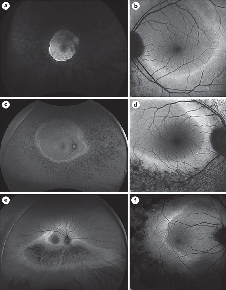
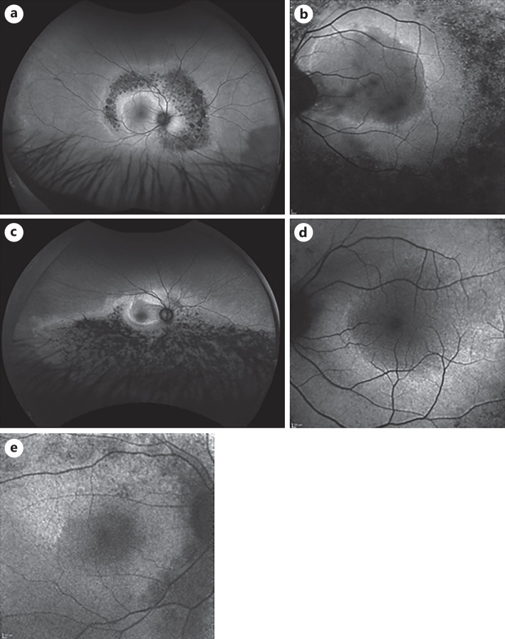
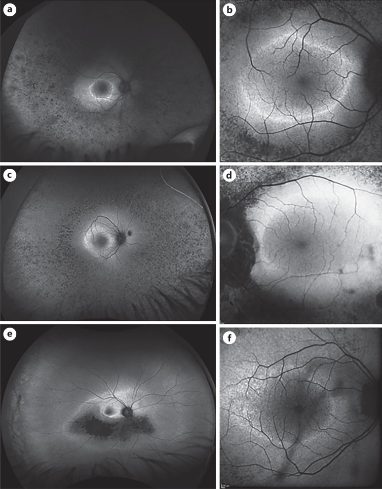
Results: Four primary fundus AF phenotypes were identified: (1) hyperAF arc, (2) hyperAF ring, (3) hyperAF ring with abnormal hyperAF within the ring, and (4) central hyperAF. The second phenotype was most common and had 3 subgroups, hyperAF rings within the macula, those extending outside the macula, and incomplete rings. HyperAF rings were also characterized as narrow or wide with wide rings having a greater amount of hyperAF. Linear Mixed-Effects Model showed mean measured EZ width was significantly greater for phenotype 1 versus each of the other 3 phenotypes (p < 0.01) and for phenotype 2 versus phenotypes 3 and 4 (p < 0.05), and also differed among phenotype 2 subgroups (p < 0.05). Other AF characteristics identified were focal posterior distinct or indistinct hypoAF which sometimes formed complete or incomplete hypoAF rings surrounding a hyperAF ring, diffuse or focal hyperAF outside hyperAF rings, and the amount of encroachment of peripheral hypoAF.
Conclusions: A grading scheme for macular AF features in patients with RP identified phenotypes that correlate with stage of disease based upon EZ width. Longitudinal studies are needed to test whether presumed early AF phenotypes evolve into later phenotypes. Use of the grading scheme for patient populations in interventional trials could help determine if any of the defined AF features provide predictive value for therapeutic responses.
期刊介绍:
''Ophthalmic Research'' features original papers and reviews reporting on translational and clinical studies. Authors from throughout the world cover research topics on every field in connection with physical, physiologic, pharmacological, biochemical and molecular biological aspects of ophthalmology. This journal also aims to provide a record of international clinical research for both researchers and clinicians in ophthalmology. Finally, the transfer of information from fundamental research to clinical research and clinical practice is particularly welcome.


 分享
分享
 求助内容:
求助内容: 应助结果提醒方式:
 扫码关注我们
