Radhika Jain, Rose Kamal, Manoj K Semwal, Deepak Thaper, Shefali Kanwar, Tripti Saxena
{"title":"Analysis of Planning Risk Volume for Heart during Radiotherapy Delivery with Breath-Hold Technique for Carcinoma of Left Breast.","authors":"Radhika Jain, Rose Kamal, Manoj K Semwal, Deepak Thaper, Shefali Kanwar, Tripti Saxena","doi":"10.4103/jmp.jmp_45_24","DOIUrl":null,"url":null,"abstract":"<p><strong>Purpose: </strong>The purpose of the study was to analyze and estimate planning risk volume (PRV) margin for heart in deep inspiration breath hold (DIBH)-based left breast radiotherapy.</p><p><strong>Materials and methods: </strong>Fifty left-sided cancer breast cases treated with volumetric modulated arc radiotherapy were included in this retrospective study. Treatment plans were created using the Eclipse treatment planning system from Varian Medical System. The treatment was delivered on TrueBeam linear accelerator (Varian). Onboard cone-beam computed tomography (CBCT) images were generated and image registration between the planning computed tomography images and the CBCT images was performed before treatment delivery. The registration provided the shifts (errors) values in 6° of freedom, namely three translational and three rotational. From the shift values, the systematic and random errors were estimated which were used to estimate PRV margin for the heart after incorporating the rotational errors with the translational errors.</p><p><strong>Results: </strong>The systematic error values after incorporating rotational errors with translational errors were 0.13 cm (lateral) and 0.11 cm (cranio caudal [CC] and anterioposterior each), and the random error values were 0.16 cm (lateral) and 0.13 cm (CC and anterioposterior each). Based on these values, the PRV margins for the heart in all three directions were 0.24 cm (lateral), 0.20 cm (CC), and 0.19 cm (anterioposterior).</p><p><strong>Conclusion: </strong>As per our institutional practice, the 2 mm value for PRV margin for the heart in all the three directions would suffice for appropriate sparing of the heart during DIBH-based radiation therapy.</p>","PeriodicalId":51719,"journal":{"name":"Journal of Medical Physics","volume":"49 4","pages":"568-573"},"PeriodicalIF":0.7000,"publicationDate":"2024-10-01","publicationTypes":"Journal Article","fieldsOfStudy":null,"isOpenAccess":false,"openAccessPdf":"https://www.ncbi.nlm.nih.gov/pmc/articles/PMC11801082/pdf/","citationCount":"0","resultStr":null,"platform":"Semanticscholar","paperid":null,"PeriodicalName":"Journal of Medical Physics","FirstCategoryId":"1085","ListUrlMain":"https://doi.org/10.4103/jmp.jmp_45_24","RegionNum":0,"RegionCategory":null,"ArticlePicture":[],"TitleCN":null,"AbstractTextCN":null,"PMCID":null,"EPubDate":"2024/12/18 0:00:00","PubModel":"Epub","JCR":"Q4","JCRName":"RADIOLOGY, NUCLEAR MEDICINE & MEDICAL IMAGING","Score":null,"Total":0}
引用次数: 0
Abstract
Purpose: The purpose of the study was to analyze and estimate planning risk volume (PRV) margin for heart in deep inspiration breath hold (DIBH)-based left breast radiotherapy.
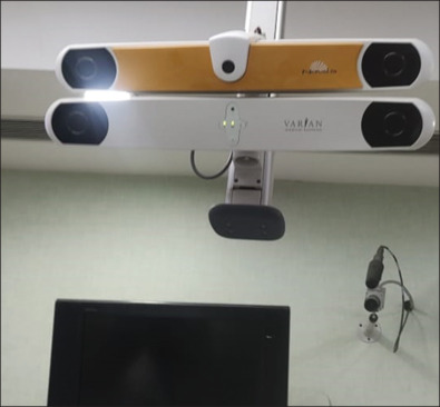
Materials and methods: Fifty left-sided cancer breast cases treated with volumetric modulated arc radiotherapy were included in this retrospective study. Treatment plans were created using the Eclipse treatment planning system from Varian Medical System. The treatment was delivered on TrueBeam linear accelerator (Varian). Onboard cone-beam computed tomography (CBCT) images were generated and image registration between the planning computed tomography images and the CBCT images was performed before treatment delivery. The registration provided the shifts (errors) values in 6° of freedom, namely three translational and three rotational. From the shift values, the systematic and random errors were estimated which were used to estimate PRV margin for the heart after incorporating the rotational errors with the translational errors.
Results: The systematic error values after incorporating rotational errors with translational errors were 0.13 cm (lateral) and 0.11 cm (cranio caudal [CC] and anterioposterior each), and the random error values were 0.16 cm (lateral) and 0.13 cm (CC and anterioposterior each). Based on these values, the PRV margins for the heart in all three directions were 0.24 cm (lateral), 0.20 cm (CC), and 0.19 cm (anterioposterior).
Conclusion: As per our institutional practice, the 2 mm value for PRV margin for the heart in all the three directions would suffice for appropriate sparing of the heart during DIBH-based radiation therapy.
期刊介绍:
JOURNAL OF MEDICAL PHYSICS is the official journal of Association of Medical Physicists of India (AMPI). The association has been bringing out a quarterly publication since 1976. Till the end of 1993, it was known as Medical Physics Bulletin, which then became Journal of Medical Physics. The main objective of the Journal is to serve as a vehicle of communication to highlight all aspects of the practice of medical radiation physics. The areas covered include all aspects of the application of radiation physics to biological sciences, radiotherapy, radiodiagnosis, nuclear medicine, dosimetry and radiation protection. Papers / manuscripts dealing with the aspects of physics related to cancer therapy / radiobiology also fall within the scope of the journal.


 分享
分享
 求助内容:
求助内容: 应助结果提醒方式:
 扫码关注我们
