Matteo Santoro, Rachel K. Lam, Sarah E. Blumenfeld, Weiqi Tan, Peter Ciari, Emily K. Chu, Nay L. Saw, Daniel Ryskamp Rijsketic, Jennifer S. Lin, Boris D. Heifets, Mehrdad Shamloo
{"title":"Mapping of catecholaminergic denervation, neurodegeneration, and inflammation in 6-OHDA-treated Parkinson’s disease mice","authors":"Matteo Santoro, Rachel K. Lam, Sarah E. Blumenfeld, Weiqi Tan, Peter Ciari, Emily K. Chu, Nay L. Saw, Daniel Ryskamp Rijsketic, Jennifer S. Lin, Boris D. Heifets, Mehrdad Shamloo","doi":"10.1038/s41531-025-00872-w","DOIUrl":null,"url":null,"abstract":"<p>Efforts to develop disease-modifying treatments for Parkinson’s disease (PD) have been hindered by the lack of animal models replicating all hallmarks of PD and the insufficient attention to extra-nigrostriatal regions pathologically critical for the prodromal appearance of non-motor symptoms. Among PD models, 6-hydroxydopamine (6-OHDA) infusion in mice has gained prominence since 2012, primarily focusing on the nigrostriatal region. This study characterized tyrosine hydroxylase-positive neuron and fiber loss across the brain following a unilateral 6-OHDA (20 µg) infusion into the dorsal striatum. Our analysis integrates immunolabeling, brain clearing (iDISCO+), light sheet microscopy, and computational methods, including fMRI and machine learning tools. We also examined sex differences, disease progression, neuroinflammatory responses, and pro-apoptotic signaling in nigrostriatal regions of C57BL/6 mice exposed to varying 6-OHDA dosages (5, 10, or 20 µg) followed by 1, 7, and 14 days of recovery. This comprehensive, spatiotemporal analysis of 6-OHDA-induced pathology was used to map the time course of neuronal degeneration and the onset of neuroinflammation.</p>","PeriodicalId":19706,"journal":{"name":"NPJ Parkinson's Disease","volume":"86 1","pages":""},"PeriodicalIF":8.2000,"publicationDate":"2025-02-11","publicationTypes":"Journal Article","fieldsOfStudy":null,"isOpenAccess":false,"openAccessPdf":"","citationCount":"0","resultStr":null,"platform":"Semanticscholar","paperid":null,"PeriodicalName":"NPJ Parkinson's Disease","FirstCategoryId":"3","ListUrlMain":"https://doi.org/10.1038/s41531-025-00872-w","RegionNum":1,"RegionCategory":"医学","ArticlePicture":[],"TitleCN":null,"AbstractTextCN":null,"PMCID":null,"EPubDate":"","PubModel":"","JCR":"Q1","JCRName":"NEUROSCIENCES","Score":null,"Total":0}
引用次数: 0
Abstract
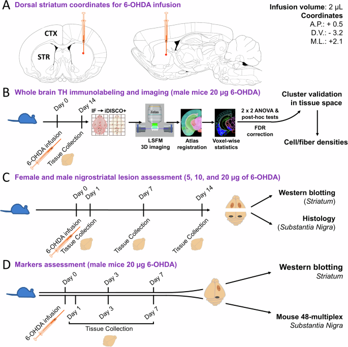
Efforts to develop disease-modifying treatments for Parkinson’s disease (PD) have been hindered by the lack of animal models replicating all hallmarks of PD and the insufficient attention to extra-nigrostriatal regions pathologically critical for the prodromal appearance of non-motor symptoms. Among PD models, 6-hydroxydopamine (6-OHDA) infusion in mice has gained prominence since 2012, primarily focusing on the nigrostriatal region. This study characterized tyrosine hydroxylase-positive neuron and fiber loss across the brain following a unilateral 6-OHDA (20 µg) infusion into the dorsal striatum. Our analysis integrates immunolabeling, brain clearing (iDISCO+), light sheet microscopy, and computational methods, including fMRI and machine learning tools. We also examined sex differences, disease progression, neuroinflammatory responses, and pro-apoptotic signaling in nigrostriatal regions of C57BL/6 mice exposed to varying 6-OHDA dosages (5, 10, or 20 µg) followed by 1, 7, and 14 days of recovery. This comprehensive, spatiotemporal analysis of 6-OHDA-induced pathology was used to map the time course of neuronal degeneration and the onset of neuroinflammation.
期刊介绍:
npj Parkinson's Disease is a comprehensive open access journal that covers a wide range of research areas related to Parkinson's disease. It publishes original studies in basic science, translational research, and clinical investigations. The journal is dedicated to advancing our understanding of Parkinson's disease by exploring various aspects such as anatomy, etiology, genetics, cellular and molecular physiology, neurophysiology, epidemiology, and therapeutic development. By providing free and immediate access to the scientific and Parkinson's disease community, npj Parkinson's Disease promotes collaboration and knowledge sharing among researchers and healthcare professionals.


 分享
分享
 求助内容:
求助内容: 应助结果提醒方式:
 扫码关注我们
