Ary Alves Mesquita-Júnior, Suelem Brenda Dos Santos, Rebeka de Oliveira Reis, Ingrid Luiza Mendonça Cunha, Aida Renée Assayag Hanan, Emílio Carlos Sponchiado-Júnior
{"title":"Endodontic Treatment of an Upper Lateral Incisor with Oehlers' Type II Dens Invaginatus: A Case Report.","authors":"Ary Alves Mesquita-Júnior, Suelem Brenda Dos Santos, Rebeka de Oliveira Reis, Ingrid Luiza Mendonça Cunha, Aida Renée Assayag Hanan, Emílio Carlos Sponchiado-Júnior","doi":"10.22037/iej.v20i1.46131","DOIUrl":null,"url":null,"abstract":"<p><p>Dens invaginatus is a developmental malformation that is caused by the invagination of the enamel organ into the internal region of the dental papilla before tissue calcification. The aim of the present report is to discuss a clinical case of endodontic treatment of tooth #12, using bioceramic sealer. The extraoral examination revealed atypical anatomy, while vitality and percussion tests were negative, palpation test was positive. Edema was observed in the adjacent gingival mucosa. Based on clinical and tomographic findings, the diagnosis was pulp necrosis with chronic periapical abscess and Oehlers' type II dens invaginatus. In the first session, access surgery was performed with spherical drills with the aid of an operating microscope (OM) and an ultrasonic diamond tip. Four canals were located, and they were partially debrided and medicated. In the second session, odontometry and chemical-mechanical preparation with nickel-titanium instruments were performed. The irrigation solution was 2.5% sodium hypochlorite. The root canals were filled with calcium hydroxide paste and the chamber was temporarily sealed. During the third session, ultrasonic irrigation was applied for final washing and the root canals were filled with Bio-C sealer using the classic single-cone technique. At 6-month follow-up, the tooth was asymptomatic and the radiography revealed significant bone repair. It was concluded that tomography, operating microscope, ultrasonic irrigation, and materials with greater flow, such as bioceramic sealers, enhanced the clinical success of the clinical case.</p>","PeriodicalId":14534,"journal":{"name":"Iranian Endodontic Journal","volume":"20 1","pages":"e7"},"PeriodicalIF":0.0000,"publicationDate":"2025-01-01","publicationTypes":"Journal Article","fieldsOfStudy":null,"isOpenAccess":false,"openAccessPdf":"https://www.ncbi.nlm.nih.gov/pmc/articles/PMC11808323/pdf/","citationCount":"0","resultStr":null,"platform":"Semanticscholar","paperid":null,"PeriodicalName":"Iranian Endodontic Journal","FirstCategoryId":"1085","ListUrlMain":"https://doi.org/10.22037/iej.v20i1.46131","RegionNum":0,"RegionCategory":null,"ArticlePicture":[],"TitleCN":null,"AbstractTextCN":null,"PMCID":null,"EPubDate":"","PubModel":"","JCR":"Q3","JCRName":"Dentistry","Score":null,"Total":0}
引用次数: 0
Abstract
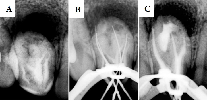
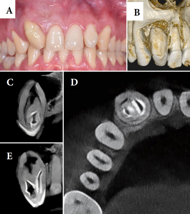
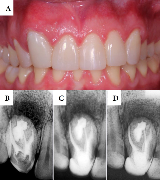
Dens invaginatus is a developmental malformation that is caused by the invagination of the enamel organ into the internal region of the dental papilla before tissue calcification. The aim of the present report is to discuss a clinical case of endodontic treatment of tooth #12, using bioceramic sealer. The extraoral examination revealed atypical anatomy, while vitality and percussion tests were negative, palpation test was positive. Edema was observed in the adjacent gingival mucosa. Based on clinical and tomographic findings, the diagnosis was pulp necrosis with chronic periapical abscess and Oehlers' type II dens invaginatus. In the first session, access surgery was performed with spherical drills with the aid of an operating microscope (OM) and an ultrasonic diamond tip. Four canals were located, and they were partially debrided and medicated. In the second session, odontometry and chemical-mechanical preparation with nickel-titanium instruments were performed. The irrigation solution was 2.5% sodium hypochlorite. The root canals were filled with calcium hydroxide paste and the chamber was temporarily sealed. During the third session, ultrasonic irrigation was applied for final washing and the root canals were filled with Bio-C sealer using the classic single-cone technique. At 6-month follow-up, the tooth was asymptomatic and the radiography revealed significant bone repair. It was concluded that tomography, operating microscope, ultrasonic irrigation, and materials with greater flow, such as bioceramic sealers, enhanced the clinical success of the clinical case.
期刊介绍:
The Iranian Endodontic Journal (IEJ) is an international peer-reviewed biomedical publication, the aim of which is to provide a scientific medium of communication for researchers throughout the globe. IEJ aims to publish the highest quality articles, both clinical and scientific, on all aspects of Endodontics. The journal is an official Journal of the Iranian Center for Endodontic Research (ICER) and the Iranian Association of Endodontists (IAE). The Journal welcomes articles related to the scientific or applied aspects of endodontics e.g. original researches, systematic reviews, meta-analyses, review articles, clinical trials, case series/reports, hypotheses, letters to the editor, etc. From the beginning (i.e. since 2006), the IEJ was the first open access endodontic journal in the world, which gave readers free and instant access to published articles and enabling them faster discovery of the latest endodontic research.


 分享
分享
 求助内容:
求助内容: 应助结果提醒方式:
 扫码关注我们
