{"title":"Bilateral Foveal Damage Induced by Indirect Picosecond Nd:YAG Laser Exposure: A Case Report.","authors":"Takahiro Miyake, Naoki Kimura, Fumi Gomi","doi":"10.1155/crop/6664488","DOIUrl":null,"url":null,"abstract":"<p><p><b>Introduction:</b> Accidental retinal injuries caused by lasers without appropriate eye protection are not rare; most cases are unilateral. We report the case of a medical nurse who sustained bilateral foveal damage through indirect exposure to a picosecond dermal laser. <b>Case Presentation:</b> A 23-year-old nurse working in a cosmetic surgery clinic was using a picosecond KTP/Nd:YAG laser for tattoo removal. Because the procedure was complicated, she neglected the use of protective eyewear and experienced dazzle. Thirty minutes after starting the procedure, she developed central scotomas in both eyes. We examined her eyes the next day. Ophthalmologic examination revealed best-corrected decimal visual acuity (BCVA) of 0.6 in the right eye and 0.3 in the left eye. Spectral domain-optical coherence tomography showed a hyperreflective inner retinal layer with a lamellar defect and focal outer retinal detachment in the right eye; the left eye exhibited intra- and subretinal foveal hemorrhages. Injections of sub-Tenon's triamcinolone acetonide (12 mg/0.3 mL) in the right eye and intravitreal tissue plasminogen activator (30 <i>μ</i>g/0.05 mL) in the left eye were administered on the same day. Two weeks later, a full-thickness macular hole (FTMH) was identified in the right eye; pars plana vitrectomy was required 6 weeks after initial presentation. Because the FTMH failed to close, a second procedure was performed 2 months later. One year after initial presentation, BCVA in the right eye had improved to 0.4. Although the FTMH remained closed, an outer retinal layer defect persisted. In the left eye, foveal hemorrhage resolved within 1 month of initial presentation. At the 1-year follow-up, BCVA in the left eye was 0.4; outer retinal layer disruption was evident at the central fovea. <b>Conclusions:</b> Continuous Nd:YAG laser exposure during cosmetic procedures likely caused the bilateral foveal damage observed in this case. All individuals using lasers must be aware of the importance of protective goggles.</p>","PeriodicalId":9603,"journal":{"name":"Case Reports in Ophthalmological Medicine","volume":"2025 ","pages":"6664488"},"PeriodicalIF":0.4000,"publicationDate":"2025-02-07","publicationTypes":"Journal Article","fieldsOfStudy":null,"isOpenAccess":false,"openAccessPdf":"https://www.ncbi.nlm.nih.gov/pmc/articles/PMC11828650/pdf/","citationCount":"0","resultStr":null,"platform":"Semanticscholar","paperid":null,"PeriodicalName":"Case Reports in Ophthalmological Medicine","FirstCategoryId":"1085","ListUrlMain":"https://doi.org/10.1155/crop/6664488","RegionNum":0,"RegionCategory":null,"ArticlePicture":[],"TitleCN":null,"AbstractTextCN":null,"PMCID":null,"EPubDate":"2025/1/1 0:00:00","PubModel":"eCollection","JCR":"Q4","JCRName":"OPHTHALMOLOGY","Score":null,"Total":0}
引用次数: 0
Abstract
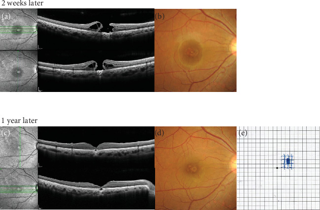
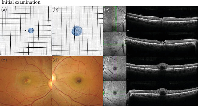
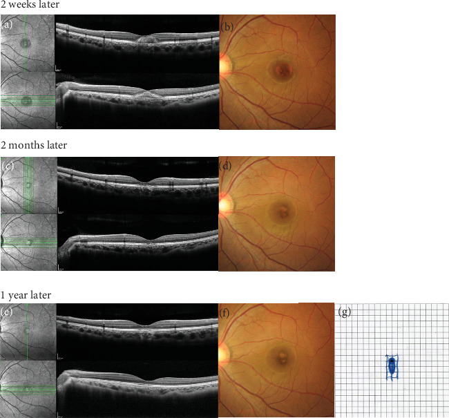
Introduction: Accidental retinal injuries caused by lasers without appropriate eye protection are not rare; most cases are unilateral. We report the case of a medical nurse who sustained bilateral foveal damage through indirect exposure to a picosecond dermal laser. Case Presentation: A 23-year-old nurse working in a cosmetic surgery clinic was using a picosecond KTP/Nd:YAG laser for tattoo removal. Because the procedure was complicated, she neglected the use of protective eyewear and experienced dazzle. Thirty minutes after starting the procedure, she developed central scotomas in both eyes. We examined her eyes the next day. Ophthalmologic examination revealed best-corrected decimal visual acuity (BCVA) of 0.6 in the right eye and 0.3 in the left eye. Spectral domain-optical coherence tomography showed a hyperreflective inner retinal layer with a lamellar defect and focal outer retinal detachment in the right eye; the left eye exhibited intra- and subretinal foveal hemorrhages. Injections of sub-Tenon's triamcinolone acetonide (12 mg/0.3 mL) in the right eye and intravitreal tissue plasminogen activator (30 μg/0.05 mL) in the left eye were administered on the same day. Two weeks later, a full-thickness macular hole (FTMH) was identified in the right eye; pars plana vitrectomy was required 6 weeks after initial presentation. Because the FTMH failed to close, a second procedure was performed 2 months later. One year after initial presentation, BCVA in the right eye had improved to 0.4. Although the FTMH remained closed, an outer retinal layer defect persisted. In the left eye, foveal hemorrhage resolved within 1 month of initial presentation. At the 1-year follow-up, BCVA in the left eye was 0.4; outer retinal layer disruption was evident at the central fovea. Conclusions: Continuous Nd:YAG laser exposure during cosmetic procedures likely caused the bilateral foveal damage observed in this case. All individuals using lasers must be aware of the importance of protective goggles.


 分享
分享
 求助内容:
求助内容: 应助结果提醒方式:
 扫码关注我们
