FAP+ activated fibroblasts are detectable in the microenvironment of endometriosis and correlate with stroma composition and infiltrating CD8+ and CD68+ cells.
Franziska Kellers, Ulf Lützen, Frederik Verburg, Annett Lebenatus, Karolin Tesch, Fatih Yalcin, Moritz Jesinghaus, Valentina Stoll, Hanna Grebe, Christoph Röcken, Dirk Bauerschlag, Björn Konukiewitz
{"title":"FAP+ activated fibroblasts are detectable in the microenvironment of endometriosis and correlate with stroma composition and infiltrating CD8+ and CD68+ cells.","authors":"Franziska Kellers, Ulf Lützen, Frederik Verburg, Annett Lebenatus, Karolin Tesch, Fatih Yalcin, Moritz Jesinghaus, Valentina Stoll, Hanna Grebe, Christoph Röcken, Dirk Bauerschlag, Björn Konukiewitz","doi":"10.1093/hropen/hoaf003","DOIUrl":null,"url":null,"abstract":"<p><strong>Study question: </strong>Do activated fibroblasts expressing fibroblast activation protein-α (FAP) - which is traceable in positron emission topography/computed topography (PET/CT) - play a role in the microenvironment of endometriosis?</p><p><strong>Summary answer: </strong>Activated fibroblasts expressing FAP are detectable in endometriotic lesions and correlate with iron and collagen content and infiltrating CD8-positive cytotoxic T cells and CD68-positive macrophages in the microenvironment endometriotic lesions.</p><p><strong>What is known already: </strong>FAP-positive activated fibroblasts are found in various fibrosis-related pathologies and in the desmoplastic stroma of solid tumours; they can be traced in PET/CT but have not been investigated in the context of endometriosis, a chronic disease involving hormone-mediated repetitive tissue remodelling and fibrosis.</p><p><strong>Study design size duration: </strong>We analysed a cohort of endometriosis patients (n = 159) who had undergone surgery with removal of endometriotic foci at our University Hospital (tertiary care centre) between 2018 and 2024. All patients provided written informed consent. The median age of the patients was 34 years. In total, 245 samples from different locations were analysed retrospectively.</p><p><strong>Participants/materials setting methods: </strong>We investigated the expression of FAP and its relation to stroma composition and the immune microenvironment of endometriosis in 245 specimens from peritoneal lesions, ovarian endometriomas, deep infiltrating endometriosis, and extra-abdominal lesions using conventional histology and immunohistochemistry followed by digital image analysis. Tissue within a radius of 500 µm of ectopic endometrium-like epithelium was analysed. To measure FAP expression in the perilesional stroma, a histoscore (H-score) was calculated. Masson trichrome staining was used to determine collagen content. Prussian blue staining for iron was used for age-dating of lesions. The abundance of CD68-positive macrophages and CD8-positive cytotoxic T cells within the microenvironment of ectopic endometriotic glands was analysed. Extra-lesional tissue served as controls.</p><p><strong>Main results and the role of chance: </strong>Distinct FAP expression (H-score >10) was observed in 84% of endometriotic lesions and in only 4% of extra-lesional controls. FAP expression was significantly higher in endometriotic lesions (mean H-score 61.8) than in extra-lesional tissue (mean H-score 3.8, <i>P</i> < 0.0001). There was a significant (<i>P</i> < 0.05) association with collagen content when comparing samples with low (H-score <100) and high (H-score ≥100) FAP expression, and a significant difference in FAP expression correlating with the tissue iron content when comparing strong staining intensity and negative samples (<i>P</i> < 0.0005) or samples with weak staining intensity (<i>P</i> < 0.005). Moreover, the abundance of CD8-positive and CD68-positive cells was significantly higher (<i>P</i> < 0.0001) in samples with high FAP expression (H-score ≥100).</p><p><strong>Large scale data: </strong>N/A.</p><p><strong>Limitations reasons for caution: </strong>This study proves the presence of FAP-positive fibroblasts in endometriosis by immunohistological methods. However, to translate targeting FAP into endometriosis diagnostics, these results have to be compared to imaging data and FAP inhibitor (FAPi) PET/CT has to be validated in a structured way on a large patient cohort. Moreover, we show that FAP expression is intertwined with the immune cell infiltrate in the microenvironment of endometriosis. To explore and understand mechanisms contributing to chronic inflammation, immune evasion, and fibrosis, more studies including more immune cell subtypes and functional experiments are needed.</p><p><strong>Wider implications of the findings: </strong>FAP-positive activated fibroblasts not only impact the immune microenvironment of endometriosis and are linked to increased macrophage and cytotoxic T-cell infiltration, as we showed, but could also provide new options for non-invasive diagnostic methods and an improvement of the diagnostic workup prior to surgery. FAPi PET/CT should be considered when exploring new diagnostic options in endometriosis.</p><p><strong>Study funding/competing interests: </strong>This work was funded by the Deutsche Forschungsgemeinschaft (DFG, German Research Foundation) 'Clinician Scientist Program in Evolutionary Medicine' (project number 413490537 to F.K.). We acknowledge financial support by Land Schleswig-Holstein within the funding programme 'Open Access Publikationsfonds'. The authors declare that they have no conflicts of interest related to this work.</p>","PeriodicalId":73264,"journal":{"name":"Human reproduction open","volume":"2025 1","pages":"hoaf003"},"PeriodicalIF":11.1000,"publicationDate":"2025-01-24","publicationTypes":"Journal Article","fieldsOfStudy":null,"isOpenAccess":false,"openAccessPdf":"https://www.ncbi.nlm.nih.gov/pmc/articles/PMC11829078/pdf/","citationCount":"0","resultStr":null,"platform":"Semanticscholar","paperid":null,"PeriodicalName":"Human reproduction open","FirstCategoryId":"1085","ListUrlMain":"https://doi.org/10.1093/hropen/hoaf003","RegionNum":0,"RegionCategory":null,"ArticlePicture":[],"TitleCN":null,"AbstractTextCN":null,"PMCID":null,"EPubDate":"2025/1/1 0:00:00","PubModel":"eCollection","JCR":"Q1","JCRName":"OBSTETRICS & GYNECOLOGY","Score":null,"Total":0}
引用次数: 0
Abstract
Study question: Do activated fibroblasts expressing fibroblast activation protein-α (FAP) - which is traceable in positron emission topography/computed topography (PET/CT) - play a role in the microenvironment of endometriosis?
Summary answer: Activated fibroblasts expressing FAP are detectable in endometriotic lesions and correlate with iron and collagen content and infiltrating CD8-positive cytotoxic T cells and CD68-positive macrophages in the microenvironment endometriotic lesions.
What is known already: FAP-positive activated fibroblasts are found in various fibrosis-related pathologies and in the desmoplastic stroma of solid tumours; they can be traced in PET/CT but have not been investigated in the context of endometriosis, a chronic disease involving hormone-mediated repetitive tissue remodelling and fibrosis.
Study design size duration: We analysed a cohort of endometriosis patients (n = 159) who had undergone surgery with removal of endometriotic foci at our University Hospital (tertiary care centre) between 2018 and 2024. All patients provided written informed consent. The median age of the patients was 34 years. In total, 245 samples from different locations were analysed retrospectively.
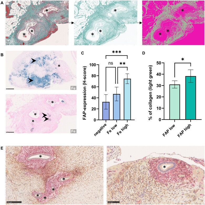
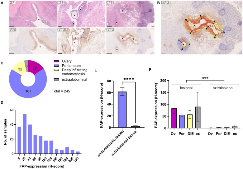
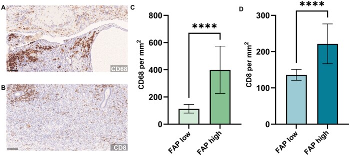
Participants/materials setting methods: We investigated the expression of FAP and its relation to stroma composition and the immune microenvironment of endometriosis in 245 specimens from peritoneal lesions, ovarian endometriomas, deep infiltrating endometriosis, and extra-abdominal lesions using conventional histology and immunohistochemistry followed by digital image analysis. Tissue within a radius of 500 µm of ectopic endometrium-like epithelium was analysed. To measure FAP expression in the perilesional stroma, a histoscore (H-score) was calculated. Masson trichrome staining was used to determine collagen content. Prussian blue staining for iron was used for age-dating of lesions. The abundance of CD68-positive macrophages and CD8-positive cytotoxic T cells within the microenvironment of ectopic endometriotic glands was analysed. Extra-lesional tissue served as controls.
Main results and the role of chance: Distinct FAP expression (H-score >10) was observed in 84% of endometriotic lesions and in only 4% of extra-lesional controls. FAP expression was significantly higher in endometriotic lesions (mean H-score 61.8) than in extra-lesional tissue (mean H-score 3.8, P < 0.0001). There was a significant (P < 0.05) association with collagen content when comparing samples with low (H-score <100) and high (H-score ≥100) FAP expression, and a significant difference in FAP expression correlating with the tissue iron content when comparing strong staining intensity and negative samples (P < 0.0005) or samples with weak staining intensity (P < 0.005). Moreover, the abundance of CD8-positive and CD68-positive cells was significantly higher (P < 0.0001) in samples with high FAP expression (H-score ≥100).
Large scale data: N/A.
Limitations reasons for caution: This study proves the presence of FAP-positive fibroblasts in endometriosis by immunohistological methods. However, to translate targeting FAP into endometriosis diagnostics, these results have to be compared to imaging data and FAP inhibitor (FAPi) PET/CT has to be validated in a structured way on a large patient cohort. Moreover, we show that FAP expression is intertwined with the immune cell infiltrate in the microenvironment of endometriosis. To explore and understand mechanisms contributing to chronic inflammation, immune evasion, and fibrosis, more studies including more immune cell subtypes and functional experiments are needed.
Wider implications of the findings: FAP-positive activated fibroblasts not only impact the immune microenvironment of endometriosis and are linked to increased macrophage and cytotoxic T-cell infiltration, as we showed, but could also provide new options for non-invasive diagnostic methods and an improvement of the diagnostic workup prior to surgery. FAPi PET/CT should be considered when exploring new diagnostic options in endometriosis.
Study funding/competing interests: This work was funded by the Deutsche Forschungsgemeinschaft (DFG, German Research Foundation) 'Clinician Scientist Program in Evolutionary Medicine' (project number 413490537 to F.K.). We acknowledge financial support by Land Schleswig-Holstein within the funding programme 'Open Access Publikationsfonds'. The authors declare that they have no conflicts of interest related to this work.
研究问题:表达成纤维细胞活化蛋白-α (FAP)的活化成纤维细胞是否在子宫内膜异位症的微环境中发挥作用? FAP可在正电子发射成像/计算机成像(PET/CT)中追踪到。总结答案:在子宫内膜异位症病变中可检测到表达FAP的活化成纤维细胞,并与微环境子宫内膜异位症病变中铁和胶原含量以及浸润的cd8阳性细胞毒性T细胞和cd68阳性巨噬细胞相关。已知情况:fap阳性活化的成纤维细胞存在于各种纤维化相关病理和实体瘤的结缔组织增生间质中;它们可以在PET/CT中追踪,但尚未在子宫内膜异位症的背景下进行研究,子宫内膜异位症是一种涉及激素介导的重复性组织重塑和纤维化的慢性疾病。研究设计规模持续时间:我们分析了一组子宫内膜异位症患者(n = 159),他们于2018年至2024年在我们的大学医院(三级护理中心)接受了子宫内膜异位症病灶切除手术。所有患者均提供书面知情同意书。患者的中位年龄为34岁。回顾性分析了来自不同地点的245份样本。研究对象/材料设置方法:采用常规组织学和免疫组织化学结合数字图像分析的方法,研究了245例腹膜病变、卵巢子宫内膜异位症、深浸润性子宫内膜异位症和腹外病变标本中FAP的表达及其与基质组成和子宫内膜异位症免疫微环境的关系。对半径为500µm的异位子宫内膜样上皮组织进行分析。为了测量FAP在病灶周围间质中的表达,计算组织评分(H-score)。马松三色染色法测定胶原蛋白含量。铁的普鲁士蓝染色用于病变的年龄测定。分析了异位子宫内膜异位症腺体微环境中cd68阳性巨噬细胞和cd8阳性细胞毒性T细胞的丰度。病变外组织作为对照。主要结果和偶发因素的作用:在84%的子宫内膜异位症病变中观察到明显的FAP表达(h评分bbb10),而在病变外对照组中仅4%。FAP在子宫内膜异位症病变(平均h评分61.8)中的表达明显高于病变外组织(平均h评分3.8,P P P P P P P)。局限性:本研究通过免疫组织学方法证实了子宫内膜异位症中fap阳性成纤维细胞的存在。然而,为了将靶向FAP转化为子宫内膜异位症的诊断,这些结果必须与成像数据和FAP抑制剂(FAPi) PET/CT进行比较,必须在大型患者队列中以结构化的方式进行验证。此外,我们发现在子宫内膜异位症的微环境中,FAP的表达与免疫细胞浸润交织在一起。为了探索和理解慢性炎症、免疫逃避和纤维化的机制,需要更多的研究,包括更多的免疫细胞亚型和功能实验。研究结果的更广泛意义:如我们所示,fap阳性激活的成纤维细胞不仅影响子宫内膜异位症的免疫微环境,与巨噬细胞和细胞毒性t细胞浸润增加有关,而且还可以为非侵入性诊断方法提供新的选择,并改善术前诊断检查。在探索子宫内膜异位症的新诊断选择时应考虑FAPi PET/CT。研究经费/竞争利益:本研究由德国研究基金会(DFG)资助。“进化医学临床科学家计划”(项目编号413490537给F.K.)。我们感谢石勒苏益格-荷尔斯泰因州在“开放获取publikationsfunds”资助计划中的财政支持。作者声明他们没有与这项工作相关的利益冲突。


 分享
分享

 求助内容:
求助内容: 应助结果提醒方式:
 扫码关注我们
