Ahmed Ibrahim, Khaled M. A. Hassanein, Shereen Ibrahim Zakaria Hussein, Mohammed M. A. Semieka and Abdelnaby M. Elshahawy
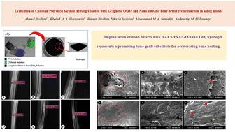
{"title":"Evaluation of a chitosan/polyvinyl alcohol hydrogel loaded with graphene oxide and nano TiO2 for bone defect reconstruction in a dog model","authors":"Ahmed Ibrahim, Khaled M. A. Hassanein, Shereen Ibrahim Zakaria Hussein, Mohammed M. A. Semieka and Abdelnaby M. Elshahawy","doi":"10.1039/D4TB02553A","DOIUrl":null,"url":null,"abstract":"<p >This study evaluated the application of chitosan/polyvinyl alcohol/graphene oxide/nano titanium oxide (CS/PVA/GO/nano TiO<small><sub>2</sub></small>) hydrogels for bone defect reconstruction in dogs. Dogs were subjected to mid-diaphyseal circular bone defects (0.8 cm<small><sup>2</sup></small>) in the radius bones. Bone defects were implanted with the hydrogel in the treated group (<em>n</em> = 9), while the control group were subjected to spontaneous healing (<em>n</em> = 9). Dogs were subjected to clinical, radiographic, and scanning electron microscopy (SEM) evaluations at 15-, 30-, and 45-days post-surgery. Dogs in the treated group recorded no lameness by the end of the third week post-surgery, while dogs in the untreated group still exhibited lameness of grade 1. There was a significant decrease (<em>p</em> < 0.05) in the cortical defect (mm) of the treated group (5.46 ± 0.17 and 1.45 ± 0.13) compared with the control group (7.57 ± 0.05 and 7.59 ± 0.06) at 30- and 45-days post-surgery, respectively. The depth of the bone defects (mm) decreased significantly (<em>p</em> < 0.05) in the treated group (2.26 ± 0.12 and 0.008 ± 0.002) compared with the untreated group (4.05 ± 0.05 and 2.16 ± 0.07) at 30- and 45-days post-surgery, respectively. Throughout the period of study, there was a significant increase (<em>p</em> < 0.05) in the radiographic density of the bone defects (px) in the treated group (474 ± 17.88) compared with that in the control group (619.6 ± 6.85). SEM results revealed complete closure of the bone defects in the treated group. Thus, implantation of bone defects with the CS/PVA/GO/nano TiO<small><sub>2</sub></small> hydrogel represents a promising bone graft substitute for accelerating bone healing.</p>","PeriodicalId":83,"journal":{"name":"Journal of Materials Chemistry B","volume":" 11","pages":" 3581-3592"},"PeriodicalIF":6.1000,"publicationDate":"2025-02-17","publicationTypes":"Journal Article","fieldsOfStudy":null,"isOpenAccess":false,"openAccessPdf":"","citationCount":"0","resultStr":null,"platform":"Semanticscholar","paperid":null,"PeriodicalName":"Journal of Materials Chemistry B","FirstCategoryId":"1","ListUrlMain":"https://pubs.rsc.org/en/content/articlelanding/2025/tb/d4tb02553a","RegionNum":3,"RegionCategory":"医学","ArticlePicture":[],"TitleCN":null,"AbstractTextCN":null,"PMCID":null,"EPubDate":"","PubModel":"","JCR":"Q1","JCRName":"MATERIALS SCIENCE, BIOMATERIALS","Score":null,"Total":0}
引用次数: 0
Abstract
This study evaluated the application of chitosan/polyvinyl alcohol/graphene oxide/nano titanium oxide (CS/PVA/GO/nano TiO2) hydrogels for bone defect reconstruction in dogs. Dogs were subjected to mid-diaphyseal circular bone defects (0.8 cm2) in the radius bones. Bone defects were implanted with the hydrogel in the treated group (n = 9), while the control group were subjected to spontaneous healing (n = 9). Dogs were subjected to clinical, radiographic, and scanning electron microscopy (SEM) evaluations at 15-, 30-, and 45-days post-surgery. Dogs in the treated group recorded no lameness by the end of the third week post-surgery, while dogs in the untreated group still exhibited lameness of grade 1. There was a significant decrease (p < 0.05) in the cortical defect (mm) of the treated group (5.46 ± 0.17 and 1.45 ± 0.13) compared with the control group (7.57 ± 0.05 and 7.59 ± 0.06) at 30- and 45-days post-surgery, respectively. The depth of the bone defects (mm) decreased significantly (p < 0.05) in the treated group (2.26 ± 0.12 and 0.008 ± 0.002) compared with the untreated group (4.05 ± 0.05 and 2.16 ± 0.07) at 30- and 45-days post-surgery, respectively. Throughout the period of study, there was a significant increase (p < 0.05) in the radiographic density of the bone defects (px) in the treated group (474 ± 17.88) compared with that in the control group (619.6 ± 6.85). SEM results revealed complete closure of the bone defects in the treated group. Thus, implantation of bone defects with the CS/PVA/GO/nano TiO2 hydrogel represents a promising bone graft substitute for accelerating bone healing.
期刊介绍:
Journal of Materials Chemistry A, B & C cover high quality studies across all fields of materials chemistry. The journals focus on those theoretical or experimental studies that report new understanding, applications, properties and synthesis of materials. Journal of Materials Chemistry A, B & C are separated by the intended application of the material studied. Broadly, applications in energy and sustainability are of interest to Journal of Materials Chemistry A, applications in biology and medicine are of interest to Journal of Materials Chemistry B, and applications in optical, magnetic and electronic devices are of interest to Journal of Materials Chemistry C.Journal of Materials Chemistry B is a Transformative Journal and Plan S compliant. Example topic areas within the scope of Journal of Materials Chemistry B are listed below. This list is neither exhaustive nor exclusive:
Antifouling coatings
Biocompatible materials
Bioelectronics
Bioimaging
Biomimetics
Biomineralisation
Bionics
Biosensors
Diagnostics
Drug delivery
Gene delivery
Immunobiology
Nanomedicine
Regenerative medicine & Tissue engineering
Scaffolds
Soft robotics
Stem cells
Therapeutic devices


 分享
分享
 求助内容:
求助内容: 应助结果提醒方式:
 扫码关注我们
