{"title":"Evaluation of <i>PPAR-α</i>, <i>PPAR-γ</i>, <i>TLR2</i>, <i>TLR4</i> Gene Expression In Patients with Coronary Artery Disease (CAD): An Experimental Study.","authors":"Mahbobe Abbasluo, Mahya Bakhshi Ardakani, Negar Jafari, Mahboubeh Pazoki","doi":"10.47176/mjiri.38.128","DOIUrl":null,"url":null,"abstract":"<p><strong>Background: </strong>Coronary artery disease (CAD) is one of the heart diseases that causes the death of many patients in the world. Many genes and molecular pathways are involved in the regulation of inflammation. However, some genes have a regulatory role and control immune responses. In recent studies, few studies have been done regarding the role of TLRs and PPARs in CAD. Hence, the present study aimed to determine and compare the mRNA expression of <i>PPAR-α</i> and <i>PPAR-γ</i> genes and genes of the innate immune system messenger pathway, including TLR2and TLR4, in CAD patients in comparison to normal individuals.</p><p><strong>Methods: </strong>This study (case-control) was conducted on 12 patients with coronary arteries and 10 healthy individuals as healthy controls. RNA extraction was performed, cDNA was produced, and then the mRNA expression levels of <i>TLR2</i>, <i>TLR4</i>, <i>PPAR</i>-α, and <i>PPAR</i>-γ genes were examined using Syber green Real-Time PCR. The t-test sample and the related non-parametric tests were used to investigate the relationship between the quantitative variables. The significance level in all tests was considered as less than 0.05.</p><p><strong>Results: </strong>The results of data analysis showed that the expression level of <i>TLR2</i> and <i>TLR4</i> genes was significantly increased in the patient group compared to the controls (<i>P</i>=0.001). However, although <i>PPAR</i>-α and <i>PPAR</i>-γ genes were up-regulated in patients' samples, the comparison of gene expression levels did not significantly differ between the case and control groups.</p><p><strong>Conclusion: </strong>we found meaningful results to the significant role of 2 and TLR4 in the pathogenesis of CAD and emphasize the hypothesis that TLR2 and TLR4 can be considered therapeutic options.</p>","PeriodicalId":18361,"journal":{"name":"Medical Journal of the Islamic Republic of Iran","volume":"38 ","pages":"128"},"PeriodicalIF":0.0000,"publicationDate":"2024-11-05","publicationTypes":"Journal Article","fieldsOfStudy":null,"isOpenAccess":false,"openAccessPdf":"https://www.ncbi.nlm.nih.gov/pmc/articles/PMC11835405/pdf/","citationCount":"0","resultStr":null,"platform":"Semanticscholar","paperid":null,"PeriodicalName":"Medical Journal of the Islamic Republic of Iran","FirstCategoryId":"1085","ListUrlMain":"https://doi.org/10.47176/mjiri.38.128","RegionNum":0,"RegionCategory":null,"ArticlePicture":[],"TitleCN":null,"AbstractTextCN":null,"PMCID":null,"EPubDate":"2024/1/1 0:00:00","PubModel":"eCollection","JCR":"Q2","JCRName":"Medicine","Score":null,"Total":0}
引用次数: 0
Abstract
Background: Coronary artery disease (CAD) is one of the heart diseases that causes the death of many patients in the world. Many genes and molecular pathways are involved in the regulation of inflammation. However, some genes have a regulatory role and control immune responses. In recent studies, few studies have been done regarding the role of TLRs and PPARs in CAD. Hence, the present study aimed to determine and compare the mRNA expression of PPAR-α and PPAR-γ genes and genes of the innate immune system messenger pathway, including TLR2and TLR4, in CAD patients in comparison to normal individuals.
Methods: This study (case-control) was conducted on 12 patients with coronary arteries and 10 healthy individuals as healthy controls. RNA extraction was performed, cDNA was produced, and then the mRNA expression levels of TLR2, TLR4, PPAR-α, and PPAR-γ genes were examined using Syber green Real-Time PCR. The t-test sample and the related non-parametric tests were used to investigate the relationship between the quantitative variables. The significance level in all tests was considered as less than 0.05.
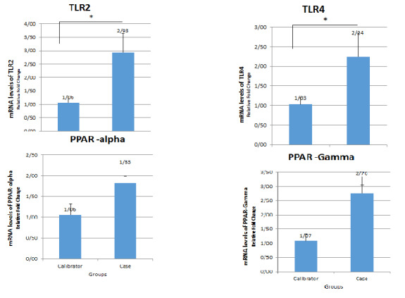
Results: The results of data analysis showed that the expression level of TLR2 and TLR4 genes was significantly increased in the patient group compared to the controls (P=0.001). However, although PPAR-α and PPAR-γ genes were up-regulated in patients' samples, the comparison of gene expression levels did not significantly differ between the case and control groups.
Conclusion: we found meaningful results to the significant role of 2 and TLR4 in the pathogenesis of CAD and emphasize the hypothesis that TLR2 and TLR4 can be considered therapeutic options.
背景:冠状动脉疾病(CAD)是世界上导致许多患者死亡的心脏疾病之一。许多基因和分子途径参与炎症的调节。然而,一些基因具有调节作用并控制免疫反应。在最近的研究中,关于tlr和ppar在CAD中的作用的研究很少。因此,本研究旨在确定和比较CAD患者与正常人相比PPAR-α和PPAR-γ基因以及先天免疫系统信使通路基因tlr2和TLR4的mRNA表达。方法:选取12例冠心病患者和10例健康人作为健康对照,采用病例对照法。提取RNA,生成cDNA,采用Syber green Real-Time PCR检测TLR2、TLR4、PPAR-α和PPAR-γ基因mRNA表达量。采用t检验样本和相关的非参数检验来考察定量变量之间的关系。所有检验的显著性水平均小于0.05。结果:资料分析结果显示,与对照组相比,患者组TLR2和TLR4基因表达水平显著升高(P=0.001)。然而,尽管PPAR-α和PPAR-γ基因在患者样本中上调,但基因表达水平在病例组和对照组之间的比较没有显著差异。结论:我们发现了2和TLR4在CAD发病机制中的重要作用,并强调了TLR2和TLR4可以考虑作为治疗选择的假设。


 分享
分享
 求助内容:
求助内容: 应助结果提醒方式:
 扫码关注我们
