Latasha Ludwig, Heather Treleaven, Arlene Khachadoorian, Brigitte Degasperi, Ingrid Walter, Deirdre Stuart, Robert A Foster, Robert Darren Wood, R Ayesha Ali, Geoffrey A Wood
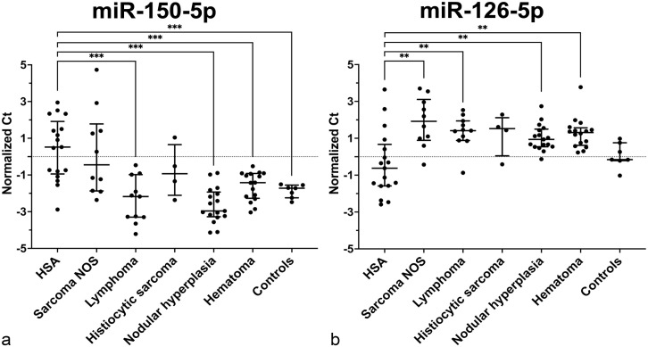
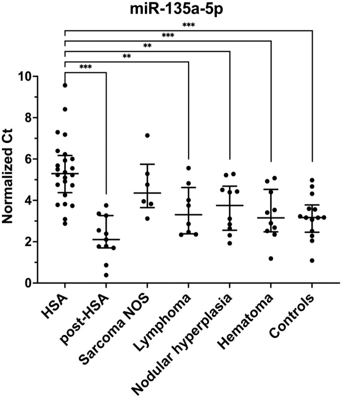
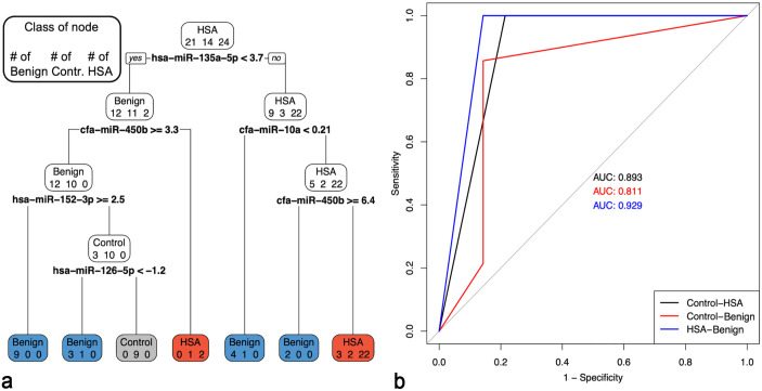
{"title":"MicroRNAs in serum and tissue can differentiate splenic hemangiosarcoma from other splenic masses in dogs.","authors":"Latasha Ludwig, Heather Treleaven, Arlene Khachadoorian, Brigitte Degasperi, Ingrid Walter, Deirdre Stuart, Robert A Foster, Robert Darren Wood, R Ayesha Ali, Geoffrey A Wood","doi":"10.1177/03009858251317466","DOIUrl":null,"url":null,"abstract":"<p><p>Splenic masses are common in dogs and vary dramatically in their clinical behavior. Clinically, and even with histology, it can be challenging to differentiate between benign and malignant splenic masses. Hemangiosarcoma (HSA), the most common malignancy of the spleen, is a very aggressive tumor with a poor prognosis. We hypothesize that microRNAs (miRNAs) in mass tissue and serum can differentiate between HSA and other splenic masses. Fifty-nine miRNAs were investigated by reverse-transcription followed by real-time quantitative polymerase chain reaction (RT-qPCR) in serum and/or tissue from dogs with HSAs (serum <i>n</i> = 24 and tissue <i>n</i> = 17; postsplenectomy serum <i>n</i> = 11), lymphomas (serum <i>n</i> = 8 and tissue <i>n</i> = 11), nonangiomatous nonlymphomatous sarcomas (serum <i>n</i> = 6 and tissue <i>n</i> = 10), histiocytic sarcomas (tissue <i>n</i> = 4), benign splenic masses (myelolipomas, nodular hyperplasia, and hematomas; total serum <i>n</i> = 21 and total tissue <i>n</i> = 35), and normal dogs (serum <i>n</i> = 14 and tissue <i>n</i> = 7). Numerous miRNAs were differentially expressed in serum and tissue of HSA cases compared to those with other splenic masses or normal spleens. In serum, our 5-miRNA model (miR-135a-5p, miR-10a, miR-450b, miR-152-3p, and miR-126-5p) accurately classified 100% (24/24) of dogs with HSA from normal dogs and those with a benign splenic mass (recall = 1 for HSA). The overall accuracy of the model was 86%. In HSA and benign splenic mass tissues, our 3-miRNA model (miR-126-5p, miR-502-3p, and miR-452-5p) accurately classified 96% of the cases. This study demonstrates the utility of miRNA models in serum and tissue for screening and diagnosis of HSA in dogs. Future studies include the evaluation of prospective and prediagnosis serum samples.</p>","PeriodicalId":23513,"journal":{"name":"Veterinary Pathology","volume":" ","pages":"659-671"},"PeriodicalIF":1.7000,"publicationDate":"2025-09-01","publicationTypes":"Journal Article","fieldsOfStudy":null,"isOpenAccess":false,"openAccessPdf":"https://www.ncbi.nlm.nih.gov/pmc/articles/PMC12314205/pdf/","citationCount":"0","resultStr":null,"platform":"Semanticscholar","paperid":null,"PeriodicalName":"Veterinary Pathology","FirstCategoryId":"97","ListUrlMain":"https://doi.org/10.1177/03009858251317466","RegionNum":2,"RegionCategory":"农林科学","ArticlePicture":[],"TitleCN":null,"AbstractTextCN":null,"PMCID":null,"EPubDate":"2025/2/19 0:00:00","PubModel":"Epub","JCR":"Q2","JCRName":"PATHOLOGY","Score":null,"Total":0}
引用次数: 0
Abstract
Splenic masses are common in dogs and vary dramatically in their clinical behavior. Clinically, and even with histology, it can be challenging to differentiate between benign and malignant splenic masses. Hemangiosarcoma (HSA), the most common malignancy of the spleen, is a very aggressive tumor with a poor prognosis. We hypothesize that microRNAs (miRNAs) in mass tissue and serum can differentiate between HSA and other splenic masses. Fifty-nine miRNAs were investigated by reverse-transcription followed by real-time quantitative polymerase chain reaction (RT-qPCR) in serum and/or tissue from dogs with HSAs (serum n = 24 and tissue n = 17; postsplenectomy serum n = 11), lymphomas (serum n = 8 and tissue n = 11), nonangiomatous nonlymphomatous sarcomas (serum n = 6 and tissue n = 10), histiocytic sarcomas (tissue n = 4), benign splenic masses (myelolipomas, nodular hyperplasia, and hematomas; total serum n = 21 and total tissue n = 35), and normal dogs (serum n = 14 and tissue n = 7). Numerous miRNAs were differentially expressed in serum and tissue of HSA cases compared to those with other splenic masses or normal spleens. In serum, our 5-miRNA model (miR-135a-5p, miR-10a, miR-450b, miR-152-3p, and miR-126-5p) accurately classified 100% (24/24) of dogs with HSA from normal dogs and those with a benign splenic mass (recall = 1 for HSA). The overall accuracy of the model was 86%. In HSA and benign splenic mass tissues, our 3-miRNA model (miR-126-5p, miR-502-3p, and miR-452-5p) accurately classified 96% of the cases. This study demonstrates the utility of miRNA models in serum and tissue for screening and diagnosis of HSA in dogs. Future studies include the evaluation of prospective and prediagnosis serum samples.
期刊介绍:
Veterinary Pathology (VET) is the premier international publication of basic and applied research involving domestic, laboratory, wildlife, marine and zoo animals, and poultry. Bridging the divide between natural and experimental diseases, the journal details the diagnostic investigations of diseases of animals; reports experimental studies on mechanisms of specific processes; provides unique insights into animal models of human disease; and presents studies on environmental and pharmaceutical hazards.


 分享
分享
 求助内容:
求助内容: 应助结果提醒方式:
 扫码关注我们
