{"title":"Surgical Resection of a Pseudoaneurysm of the First Dorsal Metatarsal Artery after Unsuccessful Embolization: A Case Report and Literature Review.","authors":"Hiroto Yasumura, Kenichi Arata, Goichi Yotsumoto, Hideyuki Satozono, Koichiro Shimoishi, Yoshihiro Fukumoto, Yuki Ogata, Tomoyuki Matsuba, Yoshiharu Soga","doi":"10.70352/scrj.cr.24-0020","DOIUrl":null,"url":null,"abstract":"<p><strong>Introduction: </strong>Aneurysms of peripheral foot arteries are extremely rare. Dorsalis pedis artery aneurysms account for 0.5% of peripheral artery aneurysms of the lower limbs. Here, we present a case of pseudoaneurysm of the first dorsal metatarsal artery of the foot and discuss the therapeutic strategy based on a literature review.</p><p><strong>Case presentation: </strong>A 76-year-old man with no history of foot trauma presented with pain and a pounding mass in the dorsum of the left foot. Echography revealed a 29 × 18 × 20 mm saccular aneurysm with to-and-fro blood flow. Contrast-enhanced computed tomography revealed an aneurysm in the first dorsal metatarsal artery. Angiography of the aneurysm revealed no arterial drainage. Embolization was subsequently performed only for the feeding artery, which was the proximal first dorsal metatarsal artery, using the 2 Target nanocoils (Stryker; Boston, MA, USA), resulting in successful occlusion. However, echography performed a few months after embolization revealed a recurrence of blood flow and enlargement of the coiled aneurysm. Nine months after embolization, the pain in the dorsum of the foot recurred. Therefore, we performed a surgical resection of the dorsal metatarsal artery aneurysm (38 × 26 × 26 mm) under general anesthesia. The first distal dorsal metatarsal artery exhibited pulsatile bleeding, and angiography of the distal dorsal metatarsal artery revealed a patent pedal arch and posterior tibial artery. Therefore, revascularization was not performed. The postoperative course was uneventful. The pathological examination indicated that the mass was a pseudoaneurysm.</p><p><strong>Conclusions: </strong>The treatments for peripheral foot artery aneurysms include observation, thrombin injection, ultrasound compression, embolization, surgical excision, and ligation. As the long-term outcomes of embolization for such aneurysms are unknown and cases are limited, surgical excision that is safe and definitive is recommended as the first-line treatment.</p>","PeriodicalId":22096,"journal":{"name":"Surgical Case Reports","volume":"11 1","pages":""},"PeriodicalIF":0.7000,"publicationDate":"2025-01-01","publicationTypes":"Journal Article","fieldsOfStudy":null,"isOpenAccess":false,"openAccessPdf":"https://www.ncbi.nlm.nih.gov/pmc/articles/PMC11835978/pdf/","citationCount":"0","resultStr":null,"platform":"Semanticscholar","paperid":null,"PeriodicalName":"Surgical Case Reports","FirstCategoryId":"1085","ListUrlMain":"https://doi.org/10.70352/scrj.cr.24-0020","RegionNum":0,"RegionCategory":null,"ArticlePicture":[],"TitleCN":null,"AbstractTextCN":null,"PMCID":null,"EPubDate":"2025/1/31 0:00:00","PubModel":"Epub","JCR":"Q4","JCRName":"SURGERY","Score":null,"Total":0}
引用次数: 0
Abstract
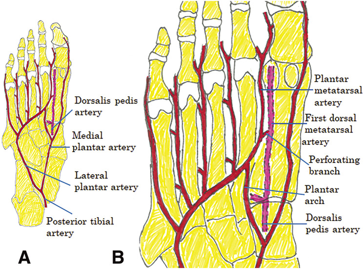
Introduction: Aneurysms of peripheral foot arteries are extremely rare. Dorsalis pedis artery aneurysms account for 0.5% of peripheral artery aneurysms of the lower limbs. Here, we present a case of pseudoaneurysm of the first dorsal metatarsal artery of the foot and discuss the therapeutic strategy based on a literature review.
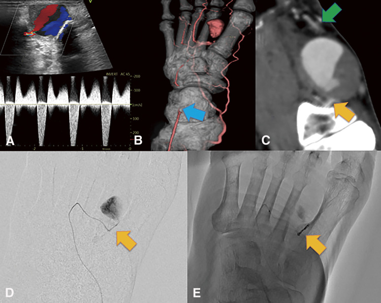
Case presentation: A 76-year-old man with no history of foot trauma presented with pain and a pounding mass in the dorsum of the left foot. Echography revealed a 29 × 18 × 20 mm saccular aneurysm with to-and-fro blood flow. Contrast-enhanced computed tomography revealed an aneurysm in the first dorsal metatarsal artery. Angiography of the aneurysm revealed no arterial drainage. Embolization was subsequently performed only for the feeding artery, which was the proximal first dorsal metatarsal artery, using the 2 Target nanocoils (Stryker; Boston, MA, USA), resulting in successful occlusion. However, echography performed a few months after embolization revealed a recurrence of blood flow and enlargement of the coiled aneurysm. Nine months after embolization, the pain in the dorsum of the foot recurred. Therefore, we performed a surgical resection of the dorsal metatarsal artery aneurysm (38 × 26 × 26 mm) under general anesthesia. The first distal dorsal metatarsal artery exhibited pulsatile bleeding, and angiography of the distal dorsal metatarsal artery revealed a patent pedal arch and posterior tibial artery. Therefore, revascularization was not performed. The postoperative course was uneventful. The pathological examination indicated that the mass was a pseudoaneurysm.
Conclusions: The treatments for peripheral foot artery aneurysms include observation, thrombin injection, ultrasound compression, embolization, surgical excision, and ligation. As the long-term outcomes of embolization for such aneurysms are unknown and cases are limited, surgical excision that is safe and definitive is recommended as the first-line treatment.


 分享
分享
 求助内容:
求助内容: 应助结果提醒方式:
 扫码关注我们
