Diogo Paula, Maria João Amaral, Joana Madeira, João Simões, André Lázaro, Nuno Silva, José Guilherme Tralhão
{"title":"Rare Encounter: A Case Report of Hepatic Perivascular Epithelioid Cell Tumor - An Uncommon Mesenchymal Tumor in the Liver.","authors":"Diogo Paula, Maria João Amaral, Joana Madeira, João Simões, André Lázaro, Nuno Silva, José Guilherme Tralhão","doi":"10.1159/000543018","DOIUrl":null,"url":null,"abstract":"<p><strong>Introduction: </strong>Perivascular epithelioid cell tumor (PEComa) is a rare neoplastic mesenchymal tumor, more frequently found in the uterus, although it can occur in different organs. Hepatic PEComa is extremely rare, with only a few cases described in the literature.</p><p><strong>Case presentation: </strong>We present a case report of a 33-year-old female patient with a history of macroprolactinoma. She was initially referred to our Department due to a 9-mm hepatic nodule incidentally diagnosed in an abdominal ultrasound in 2018. She was asymptomatic. Follow-up ultrasound showed a growth from 9 mm to 16 mm in 2019 and 30 mm in a liver magnetic resonance imaging (MRI) scan in 2022. The case was discussed in a multidisciplinary team meeting, and since malignant transformation or hepatocellular carcinoma could not be ruled out, the decision was to undergo hepatic resection. An open hepatic subsegmentectomy of segment 5 was performed, with uneventful postoperative period. The definitive diagnosis was hepatic PEComa.</p><p><strong>Conclusion: </strong>Hepatic PEComas are rare liver tumors, and their preoperative diagnosis is challenging due to the lack of specific radiological features. In most cases, the diagnosis is only confirmed through histopathological and immunohistochemical studies. Resection of the lesion appears to be the curative treatment; however, due to the rarity of the condition, there are no studies comparing surgical treatment with other options. In our case, the hypervascular lesion was initially misdiagnosed as an adenoma. PEComas should be considered as a differential diagnosis in liver nodules with well-defined margins and increased uptake in the arterial phase in computed tomography or MRI scan. Surgical resection was curative, and no recurrence was detected during the patient's follow-up.</p>","PeriodicalId":9614,"journal":{"name":"Case Reports in Gastroenterology","volume":"19 1","pages":"43-51"},"PeriodicalIF":0.6000,"publicationDate":"2025-01-24","publicationTypes":"Journal Article","fieldsOfStudy":null,"isOpenAccess":false,"openAccessPdf":"https://www.ncbi.nlm.nih.gov/pmc/articles/PMC11759453/pdf/","citationCount":"0","resultStr":null,"platform":"Semanticscholar","paperid":null,"PeriodicalName":"Case Reports in Gastroenterology","FirstCategoryId":"1085","ListUrlMain":"https://doi.org/10.1159/000543018","RegionNum":0,"RegionCategory":null,"ArticlePicture":[],"TitleCN":null,"AbstractTextCN":null,"PMCID":null,"EPubDate":"2025/1/1 0:00:00","PubModel":"eCollection","JCR":"Q4","JCRName":"GASTROENTEROLOGY & HEPATOLOGY","Score":null,"Total":0}
引用次数: 0
Abstract
Introduction: Perivascular epithelioid cell tumor (PEComa) is a rare neoplastic mesenchymal tumor, more frequently found in the uterus, although it can occur in different organs. Hepatic PEComa is extremely rare, with only a few cases described in the literature.
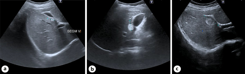
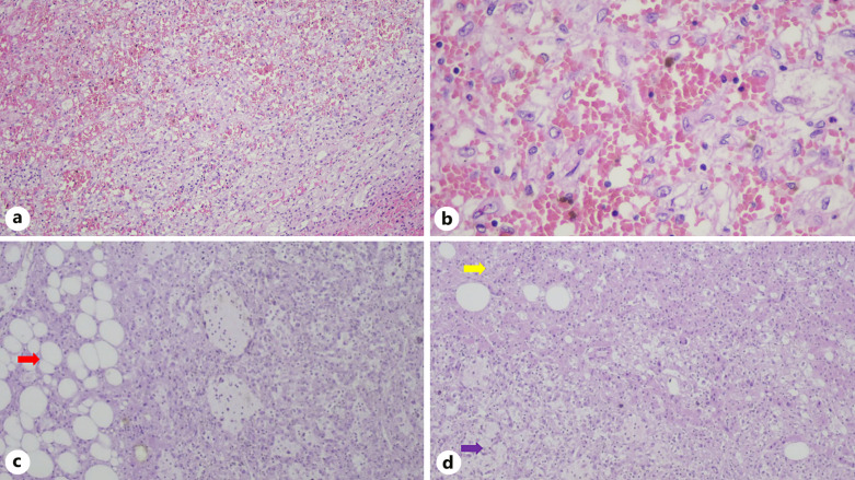
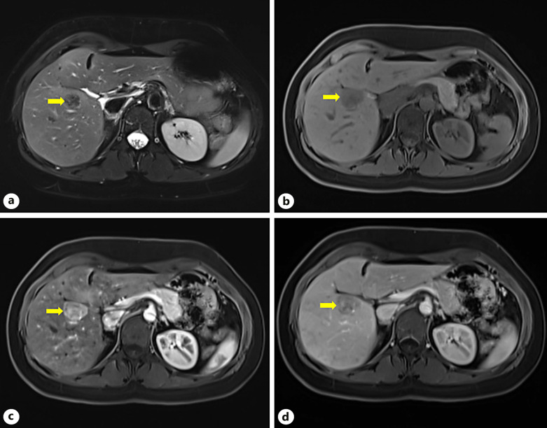
Case presentation: We present a case report of a 33-year-old female patient with a history of macroprolactinoma. She was initially referred to our Department due to a 9-mm hepatic nodule incidentally diagnosed in an abdominal ultrasound in 2018. She was asymptomatic. Follow-up ultrasound showed a growth from 9 mm to 16 mm in 2019 and 30 mm in a liver magnetic resonance imaging (MRI) scan in 2022. The case was discussed in a multidisciplinary team meeting, and since malignant transformation or hepatocellular carcinoma could not be ruled out, the decision was to undergo hepatic resection. An open hepatic subsegmentectomy of segment 5 was performed, with uneventful postoperative period. The definitive diagnosis was hepatic PEComa.
Conclusion: Hepatic PEComas are rare liver tumors, and their preoperative diagnosis is challenging due to the lack of specific radiological features. In most cases, the diagnosis is only confirmed through histopathological and immunohistochemical studies. Resection of the lesion appears to be the curative treatment; however, due to the rarity of the condition, there are no studies comparing surgical treatment with other options. In our case, the hypervascular lesion was initially misdiagnosed as an adenoma. PEComas should be considered as a differential diagnosis in liver nodules with well-defined margins and increased uptake in the arterial phase in computed tomography or MRI scan. Surgical resection was curative, and no recurrence was detected during the patient's follow-up.


 分享
分享
 求助内容:
求助内容: 应助结果提醒方式:
 扫码关注我们
