{"title":"Primary giant liposarcoma of the gallbladder: a case report and literature review.","authors":"Yu Yang, Yiwei Hou, Li Yi, Chongyuan Chen, Lihua Tang, Mingzheng Hu, Rongchun Xing","doi":"10.1186/s12957-025-03711-7","DOIUrl":null,"url":null,"abstract":"<p><strong>Background: </strong>Liposarcoma of the gallbladder is an exceptionally rare malignancy originating from adipose tissue. Its rarity and diagnostic challenges make this case noteworthy. Liposa rcomas exhibit diverse histological subtypes, each with distinct biological behaviors, and there is limited consensus on optimal treatment approaches. This report emphasizes the importance of accurate diagnosis, effective therapeutic strategies, and detailed analysis of clinical outcomes in managing such rare cases.</p><p><strong>Case presentation: </strong>A 35-year-old woman presented with a two-month history of a palpable abdominal mass accompanied by mild bloating. She reported no significant discomfort, systemic symptoms, or changes in bowel habits. Imaging revealed a large abdominal mass displacing adjacent organs. Magnetic resonance imaging suggested a mixed-signal lesion originating from the gallbladder, and laboratory tests showed elevated inflammatory markers. The patient underwent successful surgical excision of the mass and cholecystectomy. Pathological examination confirmed a well-differentiated liposarcoma closely associated with the gallbladder. Immunohistochemistry indicated positivity for CDK4, MDM2, P16, S-100, and CD34, with a low proliferation index (Ki-67 ~ 10%). Postoperative recovery was uneventful, and the patient showed significant improvement. Long-term management, including genetic testing and follow-up, was planned to monitor recurrence risk and explore potential targeted therapies.</p><p><strong>Conclusions: </strong>This case underscores the importance of considering rare malignancies like liposarcoma of the gallbladder in the differential diagnosis of abdominal masses. Early diagnosis through imaging and histopathological confirmation is crucial for optimal management. Complete surgical excision remains the cornerstone of treatment, particularly for well-differentiated subtypes, which generally have favorable prognoses. The findings highlight the need for multidisciplinary care and further research into genetic and molecular mechanisms to guide future targeted treatments.</p>","PeriodicalId":23856,"journal":{"name":"World Journal of Surgical Oncology","volume":"23 1","pages":"61"},"PeriodicalIF":2.5000,"publicationDate":"2025-02-22","publicationTypes":"Journal Article","fieldsOfStudy":null,"isOpenAccess":false,"openAccessPdf":"https://www.ncbi.nlm.nih.gov/pmc/articles/PMC11846246/pdf/","citationCount":"0","resultStr":null,"platform":"Semanticscholar","paperid":null,"PeriodicalName":"World Journal of Surgical Oncology","FirstCategoryId":"3","ListUrlMain":"https://doi.org/10.1186/s12957-025-03711-7","RegionNum":3,"RegionCategory":"医学","ArticlePicture":[],"TitleCN":null,"AbstractTextCN":null,"PMCID":null,"EPubDate":"","PubModel":"","JCR":"Q3","JCRName":"ONCOLOGY","Score":null,"Total":0}
引用次数: 0
Abstract
Background: Liposarcoma of the gallbladder is an exceptionally rare malignancy originating from adipose tissue. Its rarity and diagnostic challenges make this case noteworthy. Liposa rcomas exhibit diverse histological subtypes, each with distinct biological behaviors, and there is limited consensus on optimal treatment approaches. This report emphasizes the importance of accurate diagnosis, effective therapeutic strategies, and detailed analysis of clinical outcomes in managing such rare cases.
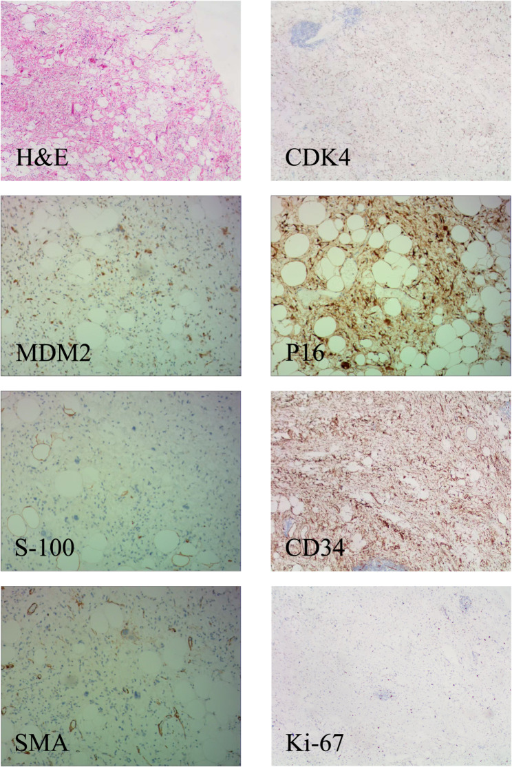
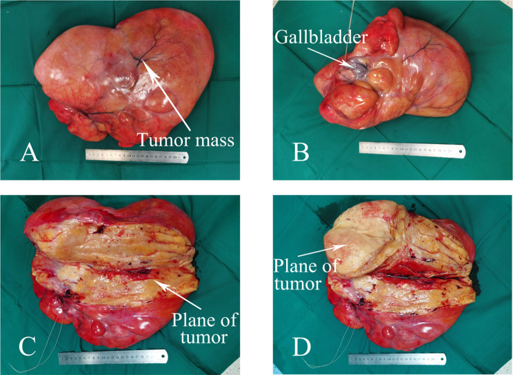
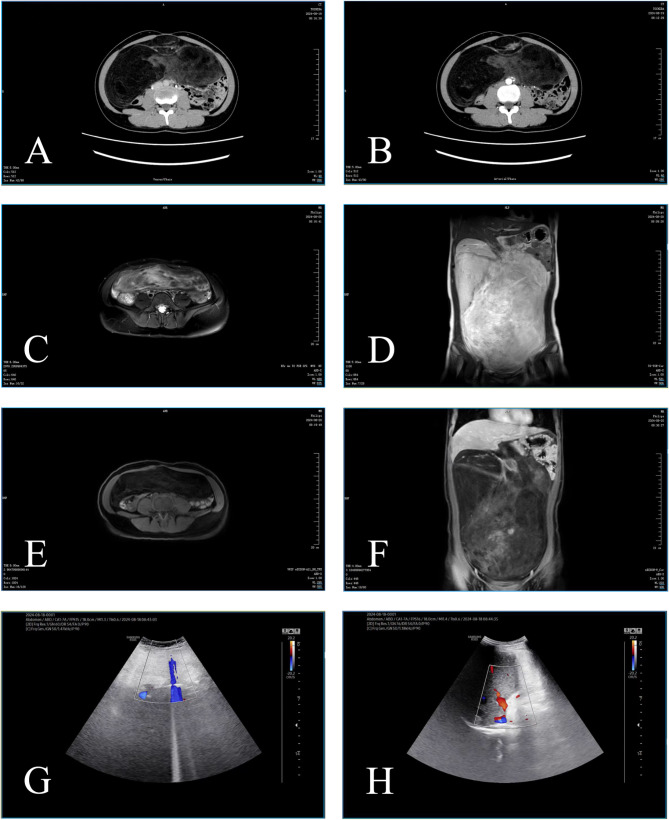
Case presentation: A 35-year-old woman presented with a two-month history of a palpable abdominal mass accompanied by mild bloating. She reported no significant discomfort, systemic symptoms, or changes in bowel habits. Imaging revealed a large abdominal mass displacing adjacent organs. Magnetic resonance imaging suggested a mixed-signal lesion originating from the gallbladder, and laboratory tests showed elevated inflammatory markers. The patient underwent successful surgical excision of the mass and cholecystectomy. Pathological examination confirmed a well-differentiated liposarcoma closely associated with the gallbladder. Immunohistochemistry indicated positivity for CDK4, MDM2, P16, S-100, and CD34, with a low proliferation index (Ki-67 ~ 10%). Postoperative recovery was uneventful, and the patient showed significant improvement. Long-term management, including genetic testing and follow-up, was planned to monitor recurrence risk and explore potential targeted therapies.
Conclusions: This case underscores the importance of considering rare malignancies like liposarcoma of the gallbladder in the differential diagnosis of abdominal masses. Early diagnosis through imaging and histopathological confirmation is crucial for optimal management. Complete surgical excision remains the cornerstone of treatment, particularly for well-differentiated subtypes, which generally have favorable prognoses. The findings highlight the need for multidisciplinary care and further research into genetic and molecular mechanisms to guide future targeted treatments.
期刊介绍:
World Journal of Surgical Oncology publishes articles related to surgical oncology and its allied subjects, such as epidemiology, cancer research, biomarkers, prevention, pathology, radiology, cancer treatment, clinical trials, multimodality treatment and molecular biology. Emphasis is placed on original research articles. The journal also publishes significant clinical case reports, as well as balanced and timely reviews on selected topics.
Oncology is a multidisciplinary super-speciality of which surgical oncology forms an integral component, especially with solid tumors. Surgical oncologists around the world are involved in research extending from detecting the mechanisms underlying the causation of cancer, to its treatment and prevention. The role of a surgical oncologist extends across the whole continuum of care. With continued developments in diagnosis and treatment, the role of a surgical oncologist is ever-changing. Hence, World Journal of Surgical Oncology aims to keep readers abreast with latest developments that will ultimately influence the work of surgical oncologists.


 分享
分享
 求助内容:
求助内容: 应助结果提醒方式:
 扫码关注我们
