{"title":"Deep-DPC: Deep learning-assisted label-free temporal imaging discovery of anti-fibrotic compounds by controlling cell morphology","authors":"Xu-dong Xing, Xiang-yu Yan, Yan-wei Tan, Yang Liu, Yi-xin Cui, Chun-ling Feng, Yu-ru Cai, Han-lin Dai, Wen Gao, Ping Zhou, Hui-ying Wang, Ping Li, Hua Yang","doi":"10.1016/j.jare.2025.02.028","DOIUrl":null,"url":null,"abstract":"<div><h3>Introduction</h3><div>Fibrosis can damage the normal function of many organs, such as cardiac function, for which no effective clinical therapies exist. However, traditional approaches to anti-fibrosis drug discovery have primarily focused on the final biological indicators, often overlooking the dynamic morphological changes during fibrosis progression. Here, we present a novel approach, <em>deep-DPC</em>, which integrates label-free, time-series digital phase contrast (DPC) imaging with cell morphology analysis and unsupervised machine learning to dynamically control and monitor cell morphology.</div></div><div><h3>Objectives</h3><div>This method enables discrimination between resting and activated fibrocytes and facilitates the discovery of non-invasive labeled anti-fibrotic lead compounds.</div></div><div><h3>Methods</h3><div>The <em>deep-DPC</em> comprises two major steps: (1) preliminary analysis by Harmony 4.9 software and (2) image classification <em>via</em> a neural network. For the experiment dataset, label-free time-series imaging was acquired from each well at 10 × magnification using the high-content imaging system, equipped with a high-speed charge-coupled device (CCD) camera. Dual-channel output images were generated through the imaging system, with one channel for bright-field and the other for DPC imaging, captured at 30-minute intervals. Firstly, applying the anti-fibrotic cell model as a case, a label-free time-series DPC imaging was developed by combining cell morphological analysis and deep learning, and its stability was verified by training with 12,000 images. Furthermore, the application of <em>deep-DPC</em> in the discovery of anti-fibrotic lead compounds.</div></div><div><h3>Results</h3><div>Using the <em>deep-DPC</em> platform, over 100,000 images generated from 1,400 compounds were processed, identifying Neo-Przewaquinone A as a potent anti-fibrosis agent. Neo-Przewaquinone A exerts its effects by inhibiting TGF-β receptor I, thereby maintaining cells in a resting state and arresting the cell cycle.</div></div><div><h3>Conclusion</h3><div>The <em>deep-DPC</em> offers a promising strategy for fibrosis assessment by combining deep learning with dynamic cell morphology analysis based on time-series DPC images. Additionally, the platform holds potential as a novel therapeutic approach for anti-myocardial fibrosis by regulating cell morphology.</div></div>","PeriodicalId":14952,"journal":{"name":"Journal of Advanced Research","volume":"78 ","pages":"Pages 703-716"},"PeriodicalIF":13.0000,"publicationDate":"2025-02-24","publicationTypes":"Journal Article","fieldsOfStudy":null,"isOpenAccess":false,"openAccessPdf":"","citationCount":"0","resultStr":null,"platform":"Semanticscholar","paperid":null,"PeriodicalName":"Journal of Advanced Research","FirstCategoryId":"103","ListUrlMain":"https://www.sciencedirect.com/science/article/pii/S2090123225001262","RegionNum":1,"RegionCategory":"综合性期刊","ArticlePicture":[],"TitleCN":null,"AbstractTextCN":null,"PMCID":null,"EPubDate":"","PubModel":"","JCR":"Q1","JCRName":"MULTIDISCIPLINARY SCIENCES","Score":null,"Total":0}
引用次数: 0
Abstract
Introduction
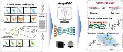
Fibrosis can damage the normal function of many organs, such as cardiac function, for which no effective clinical therapies exist. However, traditional approaches to anti-fibrosis drug discovery have primarily focused on the final biological indicators, often overlooking the dynamic morphological changes during fibrosis progression. Here, we present a novel approach, deep-DPC, which integrates label-free, time-series digital phase contrast (DPC) imaging with cell morphology analysis and unsupervised machine learning to dynamically control and monitor cell morphology.
Objectives
This method enables discrimination between resting and activated fibrocytes and facilitates the discovery of non-invasive labeled anti-fibrotic lead compounds.
Methods
The deep-DPC comprises two major steps: (1) preliminary analysis by Harmony 4.9 software and (2) image classification via a neural network. For the experiment dataset, label-free time-series imaging was acquired from each well at 10 × magnification using the high-content imaging system, equipped with a high-speed charge-coupled device (CCD) camera. Dual-channel output images were generated through the imaging system, with one channel for bright-field and the other for DPC imaging, captured at 30-minute intervals. Firstly, applying the anti-fibrotic cell model as a case, a label-free time-series DPC imaging was developed by combining cell morphological analysis and deep learning, and its stability was verified by training with 12,000 images. Furthermore, the application of deep-DPC in the discovery of anti-fibrotic lead compounds.
Results
Using the deep-DPC platform, over 100,000 images generated from 1,400 compounds were processed, identifying Neo-Przewaquinone A as a potent anti-fibrosis agent. Neo-Przewaquinone A exerts its effects by inhibiting TGF-β receptor I, thereby maintaining cells in a resting state and arresting the cell cycle.
Conclusion
The deep-DPC offers a promising strategy for fibrosis assessment by combining deep learning with dynamic cell morphology analysis based on time-series DPC images. Additionally, the platform holds potential as a novel therapeutic approach for anti-myocardial fibrosis by regulating cell morphology.
期刊介绍:
Journal of Advanced Research (J. Adv. Res.) is an applied/natural sciences, peer-reviewed journal that focuses on interdisciplinary research. The journal aims to contribute to applied research and knowledge worldwide through the publication of original and high-quality research articles in the fields of Medicine, Pharmaceutical Sciences, Dentistry, Physical Therapy, Veterinary Medicine, and Basic and Biological Sciences.
The following abstracting and indexing services cover the Journal of Advanced Research: PubMed/Medline, Essential Science Indicators, Web of Science, Scopus, PubMed Central, PubMed, Science Citation Index Expanded, Directory of Open Access Journals (DOAJ), and INSPEC.


 分享
分享
 求助内容:
求助内容: 应助结果提醒方式:
 扫码关注我们
