Luca Melazzini, Chandra Bortolotto, Leonardo Brizzi, Marina Achilli, Nicoletta Basla, Alessandro D'Onorio De Meo, Alessia Gerbasi, Olivia Maria Bottinelli, Riccardo Bellazzi, Lorenzo Preda
{"title":"AI for image quality and patient safety in CT and MRI.","authors":"Luca Melazzini, Chandra Bortolotto, Leonardo Brizzi, Marina Achilli, Nicoletta Basla, Alessandro D'Onorio De Meo, Alessia Gerbasi, Olivia Maria Bottinelli, Riccardo Bellazzi, Lorenzo Preda","doi":"10.1186/s41747-025-00562-5","DOIUrl":null,"url":null,"abstract":"<p><p>Substantial endeavors have been recently dedicated to developing artificial intelligence (AI) solutions, especially deep learning-based, tailored to enhance radiological procedures, in particular algorithms designed to minimize radiation exposure and enhance image clarity. Thus, not only better diagnostic accuracy but also reduced potential harm to patients was pursued, thereby exemplifying the intersection of technological innovation and the highest standards of patient care. We provide herein an overview of recent AI developments in computed tomography and magnetic resonance imaging. Major AI results in CT regard: optimization of patient positioning, scan range selection (avoiding \"overscanning\"), and choice of technical parameters; reduction of the amount of injected contrast agent and injection flow rate (also avoiding extravasation); faster and better image reconstruction reducing noise level and artifacts. Major AI results in MRI regard: reconstruction of undersampled images; artifact removal, including those derived from unintentional patient's (or fetal) movement or from heart motion; up to 80-90% reduction of GBCA dose. Challenges include limited generalizability, lack of external validation, insufficient explainability of models, and opacity of decision-making. Developing explainable AI algorithms that provide transparent and interpretable outputs is essential to enable seamless AI integration into CT and MRI practice. RELEVANCE STATEMENT: This review highlights how AI-driven advancements in CT and MRI improve image quality and enhance patient safety by leveraging AI solutions for dose reduction, contrast optimization, noise reduction, and efficient image reconstruction, paving the way for safer, faster, and more accurate diagnostic imaging practices. KEY POINTS: Advancements in AI are revolutionizing the way radiological images are acquired, reconstructed, and interpreted. AI algorithms can assist in optimizing radiation doses, reducing scan times, and enhancing image quality. AI techniques are paving the way for a future of more efficient, accurate, and safe medical imaging examinations.</p>","PeriodicalId":36926,"journal":{"name":"European Radiology Experimental","volume":"9 1","pages":"28"},"PeriodicalIF":3.6000,"publicationDate":"2025-02-23","publicationTypes":"Journal Article","fieldsOfStudy":null,"isOpenAccess":false,"openAccessPdf":"https://www.ncbi.nlm.nih.gov/pmc/articles/PMC11847764/pdf/","citationCount":"0","resultStr":null,"platform":"Semanticscholar","paperid":null,"PeriodicalName":"European Radiology Experimental","FirstCategoryId":"1085","ListUrlMain":"https://doi.org/10.1186/s41747-025-00562-5","RegionNum":0,"RegionCategory":null,"ArticlePicture":[],"TitleCN":null,"AbstractTextCN":null,"PMCID":null,"EPubDate":"","PubModel":"","JCR":"Q1","JCRName":"RADIOLOGY, NUCLEAR MEDICINE & MEDICAL IMAGING","Score":null,"Total":0}
引用次数: 0
Abstract
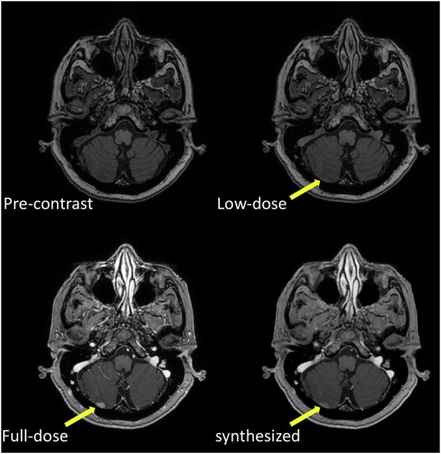
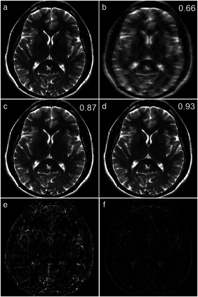
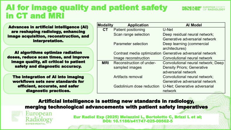
Substantial endeavors have been recently dedicated to developing artificial intelligence (AI) solutions, especially deep learning-based, tailored to enhance radiological procedures, in particular algorithms designed to minimize radiation exposure and enhance image clarity. Thus, not only better diagnostic accuracy but also reduced potential harm to patients was pursued, thereby exemplifying the intersection of technological innovation and the highest standards of patient care. We provide herein an overview of recent AI developments in computed tomography and magnetic resonance imaging. Major AI results in CT regard: optimization of patient positioning, scan range selection (avoiding "overscanning"), and choice of technical parameters; reduction of the amount of injected contrast agent and injection flow rate (also avoiding extravasation); faster and better image reconstruction reducing noise level and artifacts. Major AI results in MRI regard: reconstruction of undersampled images; artifact removal, including those derived from unintentional patient's (or fetal) movement or from heart motion; up to 80-90% reduction of GBCA dose. Challenges include limited generalizability, lack of external validation, insufficient explainability of models, and opacity of decision-making. Developing explainable AI algorithms that provide transparent and interpretable outputs is essential to enable seamless AI integration into CT and MRI practice. RELEVANCE STATEMENT: This review highlights how AI-driven advancements in CT and MRI improve image quality and enhance patient safety by leveraging AI solutions for dose reduction, contrast optimization, noise reduction, and efficient image reconstruction, paving the way for safer, faster, and more accurate diagnostic imaging practices. KEY POINTS: Advancements in AI are revolutionizing the way radiological images are acquired, reconstructed, and interpreted. AI algorithms can assist in optimizing radiation doses, reducing scan times, and enhancing image quality. AI techniques are paving the way for a future of more efficient, accurate, and safe medical imaging examinations.


 分享
分享
 求助内容:
求助内容: 应助结果提醒方式:
 扫码关注我们
