Incidentally Discovered Duodenal Gastrointestinal Stromal Tumour (GIST): Operative Treatment and Problems After Surgery-A Case Report and Literature Review.
{"title":"Incidentally Discovered Duodenal Gastrointestinal Stromal Tumour (GIST): Operative Treatment and Problems After Surgery-A Case Report and Literature Review.","authors":"Peter Lüthje, Ilona Nurmi-Lüthje","doi":"10.1155/crgm/5493240","DOIUrl":null,"url":null,"abstract":"<p><p><b>Background:</b> Gastrointestinal stromal tumours (GISTs) are mesenchymal tumours of the digestive tract that can involve any part of the tract. The tumours can be harmless or life-threatening. <b>Materials and Methods:</b> A case report of a surgeon who fell in a Finnish sauna, and he immediately felt that some ribs were broken. Magnetic resonance imaging and ultrasound showed three fractured ribs and an intrasplenic haematoma. Contrast-enhanced computed tomography (CT) demonstrated a small intrasplenic anomaly but no haematoma. Incidentally, an incidentaloma in the left adrenal gland was diagnosed. Three months later, a control CT scan was performed. The radiological findings on the adrenal gland and laboratory examinations matched those of a benign adenoma. Incidentally, a small duodenal tumour was diagnosed. At the same time, anaemia (haemoglobin: 104 g/L) and iron deficiency (ferritin: 8 μg/L) were noticed. An esophagogastroduodenoscopy showed an intramural tumour localised after the bulb-descending junction. Because the tumour was submucosal, the pathological diagnosis failed. Three months later, a radical surgical resection of the tumour with a resection margin of 2 mm and primary closing of the duodenum was performed. Pathological examination showed a well-circumscribed submucosal mesenchymal tumour with spindle cells. A tumour-free margin was uncertain. Immunohistochemistry findings showed a GIST. Due to the uncertain margin, an esophagogastroduodenoscopy control was planned at 2 years postoperatively. The patient disagreed with the decision and ordered a private control CT 3 months after the operation. The new CT found no local recurrence or metastasis. The patient contacted the head surgeon of the clinic, who ordered a 1-year postoperative CT. The 1-year follow-up CT finding agreed with the previous findings. <b>Conclusion:</b> The aftertreatment of a radical-operated GIST is extremely important if histologic examination of the tumour-free margin is uncertain. In that case, CT controls should be considered once a year for at least 3 years.</p>","PeriodicalId":45645,"journal":{"name":"Case Reports in Gastrointestinal Medicine","volume":"2025 ","pages":"5493240"},"PeriodicalIF":0.5000,"publicationDate":"2025-02-14","publicationTypes":"Journal Article","fieldsOfStudy":null,"isOpenAccess":false,"openAccessPdf":"https://www.ncbi.nlm.nih.gov/pmc/articles/PMC11845264/pdf/","citationCount":"0","resultStr":null,"platform":"Semanticscholar","paperid":null,"PeriodicalName":"Case Reports in Gastrointestinal Medicine","FirstCategoryId":"1085","ListUrlMain":"https://doi.org/10.1155/crgm/5493240","RegionNum":0,"RegionCategory":null,"ArticlePicture":[],"TitleCN":null,"AbstractTextCN":null,"PMCID":null,"EPubDate":"2025/1/1 0:00:00","PubModel":"eCollection","JCR":"Q4","JCRName":"GASTROENTEROLOGY & HEPATOLOGY","Score":null,"Total":0}
引用次数: 0
Abstract
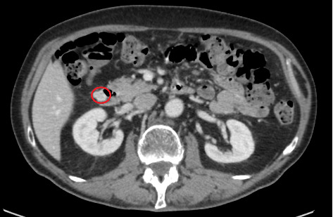
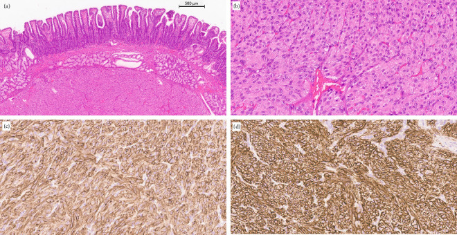
Background: Gastrointestinal stromal tumours (GISTs) are mesenchymal tumours of the digestive tract that can involve any part of the tract. The tumours can be harmless or life-threatening. Materials and Methods: A case report of a surgeon who fell in a Finnish sauna, and he immediately felt that some ribs were broken. Magnetic resonance imaging and ultrasound showed three fractured ribs and an intrasplenic haematoma. Contrast-enhanced computed tomography (CT) demonstrated a small intrasplenic anomaly but no haematoma. Incidentally, an incidentaloma in the left adrenal gland was diagnosed. Three months later, a control CT scan was performed. The radiological findings on the adrenal gland and laboratory examinations matched those of a benign adenoma. Incidentally, a small duodenal tumour was diagnosed. At the same time, anaemia (haemoglobin: 104 g/L) and iron deficiency (ferritin: 8 μg/L) were noticed. An esophagogastroduodenoscopy showed an intramural tumour localised after the bulb-descending junction. Because the tumour was submucosal, the pathological diagnosis failed. Three months later, a radical surgical resection of the tumour with a resection margin of 2 mm and primary closing of the duodenum was performed. Pathological examination showed a well-circumscribed submucosal mesenchymal tumour with spindle cells. A tumour-free margin was uncertain. Immunohistochemistry findings showed a GIST. Due to the uncertain margin, an esophagogastroduodenoscopy control was planned at 2 years postoperatively. The patient disagreed with the decision and ordered a private control CT 3 months after the operation. The new CT found no local recurrence or metastasis. The patient contacted the head surgeon of the clinic, who ordered a 1-year postoperative CT. The 1-year follow-up CT finding agreed with the previous findings. Conclusion: The aftertreatment of a radical-operated GIST is extremely important if histologic examination of the tumour-free margin is uncertain. In that case, CT controls should be considered once a year for at least 3 years.


 分享
分享
 求助内容:
求助内容: 应助结果提醒方式:
 扫码关注我们
