The Usefulness of Intraoperative Indocyanine Green Fluorescence Imaging in Surgical Treatment of Refractory Chylothorax in Pediatric Patients: A Case Report.
Kanji Ishizu, Kanta Araki, Koji Kagisaki, Hideto Ozawa
{"title":"The Usefulness of Intraoperative Indocyanine Green Fluorescence Imaging in Surgical Treatment of Refractory Chylothorax in Pediatric Patients: A Case Report.","authors":"Kanji Ishizu, Kanta Araki, Koji Kagisaki, Hideto Ozawa","doi":"10.70352/scrj.cr.24-0112","DOIUrl":null,"url":null,"abstract":"<p><strong>Introduction: </strong>Chylothorax is one of the complications in cardiovascular surgery. Although prolonged chylothorax leads to critical status and is associated with high mortality, its treatment has not been well established. We present a successful case of surgical treatment of chylothorax in a neonate using indocyanine green to identify the site of lymphatic leakage.</p><p><strong>Case presentation: </strong>The patient with complete atrioventricular septal defect, patent ductus arteriosus, pulmonary hypertension, and chromosomal abnormality with trisomy 21 underwent pulmonary artery banding and patent ductus arteriosus ligation through median sternotomy. The postoperative course was complicated with chylothorax; conservative treatment was not effective, so surgical treatment was selected. Indocyanine green was injected subcutaneously between the first and second toes on the left side 30 min before surgery to identify the site of leakage. We could detect the lymphatic leakage from the para-aortic lymph node by indocyanine green camera in the left thoracic cavity, and the leakage sites could be closed with interrupted sutures.</p><p><strong>Conclusion: </strong>Identification of lymphatic leakage sites using indocyanine green could be an effective technique in the surgical treatment of chylothorax in pediatric patients.</p>","PeriodicalId":22096,"journal":{"name":"Surgical Case Reports","volume":"11 1","pages":""},"PeriodicalIF":0.7000,"publicationDate":"2025-01-01","publicationTypes":"Journal Article","fieldsOfStudy":null,"isOpenAccess":false,"openAccessPdf":"https://www.ncbi.nlm.nih.gov/pmc/articles/PMC11850031/pdf/","citationCount":"0","resultStr":null,"platform":"Semanticscholar","paperid":null,"PeriodicalName":"Surgical Case Reports","FirstCategoryId":"1085","ListUrlMain":"https://doi.org/10.70352/scrj.cr.24-0112","RegionNum":0,"RegionCategory":null,"ArticlePicture":[],"TitleCN":null,"AbstractTextCN":null,"PMCID":null,"EPubDate":"2025/2/18 0:00:00","PubModel":"Epub","JCR":"Q4","JCRName":"SURGERY","Score":null,"Total":0}
引用次数: 0
Abstract
Introduction: Chylothorax is one of the complications in cardiovascular surgery. Although prolonged chylothorax leads to critical status and is associated with high mortality, its treatment has not been well established. We present a successful case of surgical treatment of chylothorax in a neonate using indocyanine green to identify the site of lymphatic leakage.
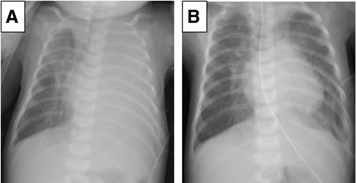
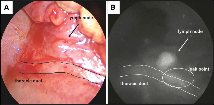
Case presentation: The patient with complete atrioventricular septal defect, patent ductus arteriosus, pulmonary hypertension, and chromosomal abnormality with trisomy 21 underwent pulmonary artery banding and patent ductus arteriosus ligation through median sternotomy. The postoperative course was complicated with chylothorax; conservative treatment was not effective, so surgical treatment was selected. Indocyanine green was injected subcutaneously between the first and second toes on the left side 30 min before surgery to identify the site of leakage. We could detect the lymphatic leakage from the para-aortic lymph node by indocyanine green camera in the left thoracic cavity, and the leakage sites could be closed with interrupted sutures.
Conclusion: Identification of lymphatic leakage sites using indocyanine green could be an effective technique in the surgical treatment of chylothorax in pediatric patients.


 分享
分享
 求助内容:
求助内容: 应助结果提醒方式:
 扫码关注我们
