Yun Wang, Christopher G Scott, Garvan C Kane, Sorin V Pislaru, Jared G Bird, Patricia A Pellikka, Vidhu Anand
{"title":"One size doesn't fit all: exploring the influence of body size, age, and sex on right ventricle size measurements.","authors":"Yun Wang, Christopher G Scott, Garvan C Kane, Sorin V Pislaru, Jared G Bird, Patricia A Pellikka, Vidhu Anand","doi":"10.1186/s13089-025-00407-7","DOIUrl":null,"url":null,"abstract":"<p><strong>Background: </strong>The assessment of right ventricular (RV) size is an important part of 2-dimensional transthoracic echocardiography. Current chamber quantification guidelines provide reference values as unindexed numbers, similar for men and women. We sought to evaluate normal ranges of RV dimensions based on age, sex, body surface area (BSA), and height. Consecutive patients with \"normal echocardiogram\" between January 2011 and August 2022 at our center were retrospectively included. RV dimensions including diameter at the base and mid-ventricle level, and base-to-apex length were measured.</p><p><strong>Results: </strong>Of 1389 patients (median 43 years, 53% female) with all three measurements available, the median RV measurements, both unindexed and indexed to BSA, were: basal diameter 35.0 mm (31.0-39.0) and 18.4 mm/m<sup>2</sup> (16.5-20.3); mid diameter 28.0 mm and 14.8 mm/m<sup>2</sup> (13.1-16.6); RV length 73.0 mm (67.0-78.0) and 37.6 mm/m<sup>2</sup> (34.9-40.9). RV dimensions were larger in men than women across all age groups but similar when indexed to BSA (for basal and mid dimensions). RV length was best indexed to height. Our indexed normal values by age and sex were similar to World Alliance Societies of Echocardiography (WASE) cohort.</p><p><strong>Conclusions: </strong>RV measurements should be indexed to BSA, considering sex and age, to determine RV size and enlargement.</p>","PeriodicalId":36911,"journal":{"name":"Ultrasound Journal","volume":"17 1","pages":"14"},"PeriodicalIF":2.9000,"publicationDate":"2025-02-24","publicationTypes":"Journal Article","fieldsOfStudy":null,"isOpenAccess":false,"openAccessPdf":"https://www.ncbi.nlm.nih.gov/pmc/articles/PMC11850662/pdf/","citationCount":"0","resultStr":null,"platform":"Semanticscholar","paperid":null,"PeriodicalName":"Ultrasound Journal","FirstCategoryId":"1085","ListUrlMain":"https://doi.org/10.1186/s13089-025-00407-7","RegionNum":0,"RegionCategory":null,"ArticlePicture":[],"TitleCN":null,"AbstractTextCN":null,"PMCID":null,"EPubDate":"","PubModel":"","JCR":"Q2","JCRName":"Medicine","Score":null,"Total":0}
引用次数: 0
Abstract
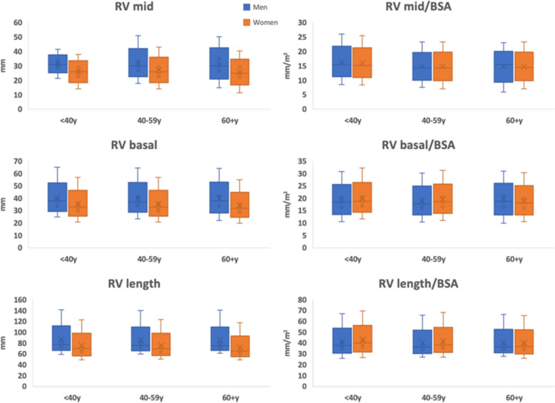
Background: The assessment of right ventricular (RV) size is an important part of 2-dimensional transthoracic echocardiography. Current chamber quantification guidelines provide reference values as unindexed numbers, similar for men and women. We sought to evaluate normal ranges of RV dimensions based on age, sex, body surface area (BSA), and height. Consecutive patients with "normal echocardiogram" between January 2011 and August 2022 at our center were retrospectively included. RV dimensions including diameter at the base and mid-ventricle level, and base-to-apex length were measured.
Results: Of 1389 patients (median 43 years, 53% female) with all three measurements available, the median RV measurements, both unindexed and indexed to BSA, were: basal diameter 35.0 mm (31.0-39.0) and 18.4 mm/m2 (16.5-20.3); mid diameter 28.0 mm and 14.8 mm/m2 (13.1-16.6); RV length 73.0 mm (67.0-78.0) and 37.6 mm/m2 (34.9-40.9). RV dimensions were larger in men than women across all age groups but similar when indexed to BSA (for basal and mid dimensions). RV length was best indexed to height. Our indexed normal values by age and sex were similar to World Alliance Societies of Echocardiography (WASE) cohort.
Conclusions: RV measurements should be indexed to BSA, considering sex and age, to determine RV size and enlargement.


 分享
分享
 求助内容:
求助内容: 应助结果提醒方式:
 扫码关注我们
