{"title":"Plaque shift to the brachiocephalic artery after subclavian artery stenting: illustrative case.","authors":"Junji Fukumori, Kenji Fukutome, Shuta Aketa, Yuki Shiraishi, Atsuko Shimotsuma, Ryuta Matsuoka, Rinsei Tei, Yasushi Shin, Yasushi Motoyama","doi":"10.3171/CASE24760","DOIUrl":null,"url":null,"abstract":"<p><strong>Background: </strong>Subclavian artery stenosis (SAS) has a prevalence of 1.9% in the general United States population. Revascularization, often by stenting, is indicated for symptomatic patients. Plaque shift (PS) is a well-known poststenting complication in coronary interventions but has not been reported in subclavian artery (SA) stenting. This case report documents the occurrence of PS after stenting for SAS, highlighting a rare but significant complication.</p><p><strong>Observations: </strong>An 87-year-old woman with a history of hypertension and dyslipidemia presented with right upper-limb pain and fatigue. Imaging confirmed stenosis with calcified plaque at the origin of the right SA. Following endovascular stenting under local anesthesia, imaging revealed PS to the brachiocephalic artery (BA). To prevent migration into the common carotid artery (CCA), a dual-layer stent was placed from the CCA to the BA. The patient's symptoms resolved, and follow-up confirmed successful plaque stabilization without restenosis 1 year postprocedure.</p><p><strong>Lessons: </strong>Even apparently hard SA plaque with calcification can result in PS with stenting. When PS occurs, prompt stenting can prevent cerebral embolism. https://thejns.org/doi/10.3171/CASE24760.</p>","PeriodicalId":94098,"journal":{"name":"Journal of neurosurgery. Case lessons","volume":"9 8","pages":""},"PeriodicalIF":0.0000,"publicationDate":"2025-02-24","publicationTypes":"Journal Article","fieldsOfStudy":null,"isOpenAccess":false,"openAccessPdf":"https://www.ncbi.nlm.nih.gov/pmc/articles/PMC12327485/pdf/","citationCount":"0","resultStr":null,"platform":"Semanticscholar","paperid":null,"PeriodicalName":"Journal of neurosurgery. Case lessons","FirstCategoryId":"1085","ListUrlMain":"https://doi.org/10.3171/CASE24760","RegionNum":0,"RegionCategory":null,"ArticlePicture":[],"TitleCN":null,"AbstractTextCN":null,"PMCID":null,"EPubDate":"","PubModel":"","JCR":"","JCRName":"","Score":null,"Total":0}
引用次数: 0
Abstract
Background: Subclavian artery stenosis (SAS) has a prevalence of 1.9% in the general United States population. Revascularization, often by stenting, is indicated for symptomatic patients. Plaque shift (PS) is a well-known poststenting complication in coronary interventions but has not been reported in subclavian artery (SA) stenting. This case report documents the occurrence of PS after stenting for SAS, highlighting a rare but significant complication.
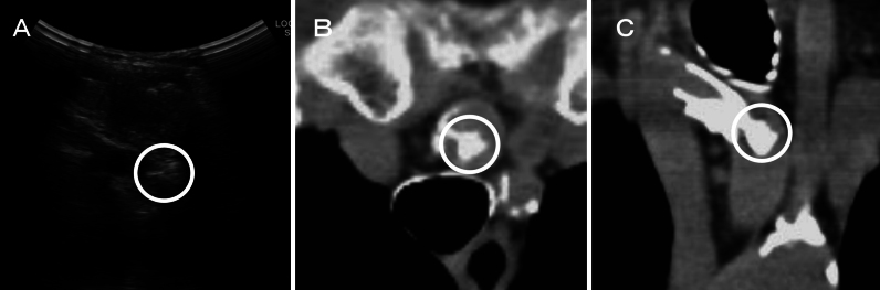
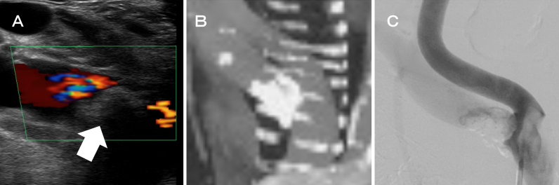
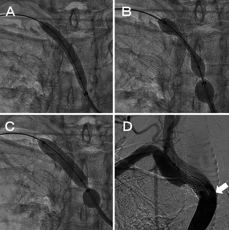
Observations: An 87-year-old woman with a history of hypertension and dyslipidemia presented with right upper-limb pain and fatigue. Imaging confirmed stenosis with calcified plaque at the origin of the right SA. Following endovascular stenting under local anesthesia, imaging revealed PS to the brachiocephalic artery (BA). To prevent migration into the common carotid artery (CCA), a dual-layer stent was placed from the CCA to the BA. The patient's symptoms resolved, and follow-up confirmed successful plaque stabilization without restenosis 1 year postprocedure.
Lessons: Even apparently hard SA plaque with calcification can result in PS with stenting. When PS occurs, prompt stenting can prevent cerebral embolism. https://thejns.org/doi/10.3171/CASE24760.


 分享
分享

 求助内容:
求助内容: 应助结果提醒方式:
 扫码关注我们
